
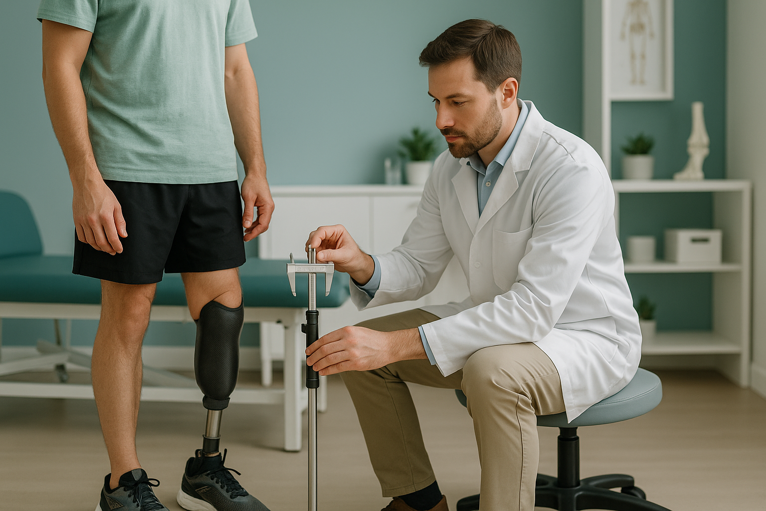
How Do Orthotists and Prosthetists Use Alignment Data?
Orthotists and prosthetists use standing whole-body alignment data to prescribe orthotics, braces, shoe raises and prosthetic components with millimetre accuracy. An EOS scan captures the full skeleton under load, from head to feet, in one image. This gives you true bone measurements, not clinical estimates. So every prescription starts from verified data.
Key takeaways
- Standing EOS imaging shows bones and joints under real weight-bearing load, not relaxed on a table.
- Reports include true bone lengths, pelvic obliquity and mechanical axis data for each limb.
- Millimetre-level accuracy supports shoe raise decisions, AFO prescription and prosthetic socket alignment.
- Before and after comparison scans let you check the effect of your intervention objectively.
- EOS uses about 90% less radiation than a standard X-ray, around ten times less.
As an orthotist or prosthetist, you already know that a few millimetres can change how a patient walks, stands and feels. Clinical assessment gives you a strong starting point. But the numbers behind a prescription need to be accurate. That is where standing whole-body imaging fits into your workflow.
Why does standing imaging matter for orthotics and prosthetics?
Standing imaging matters because your patient's alignment changes under load. When someone lies down for a conventional X-ray, CT or MRI, gravity is taken away. The pelvis levels out, curves relax and compensatory patterns disappear. You lose exactly the information you need to prescribe accurately.
The body is a connected chain from feet to head. A short leg tilts the pelvis, the spine compensates with a curve, and the shoulders follow. If your imaging shows only one region, or shows it lying down, you may miss the mechanical cause of your patient's problem.
Full weight-bearing capture. The EOS scan images the whole skeleton in one upright exposure. Pelvic obliquity, true leg lengths and mechanical axes are all measured as the patient actually loads their joints. This is the position they stand and walk in, so the data matches real life.
Muscles relax, alignment shifts. Lying-down imaging takes gravity away, so soft tissue settles and joints decompress. A leg length discrepancy may look smaller or disappear completely. Compensatory spinal curves can flatten. The numbers you take from a supine image may not reflect the alignment your patient actually lives with.
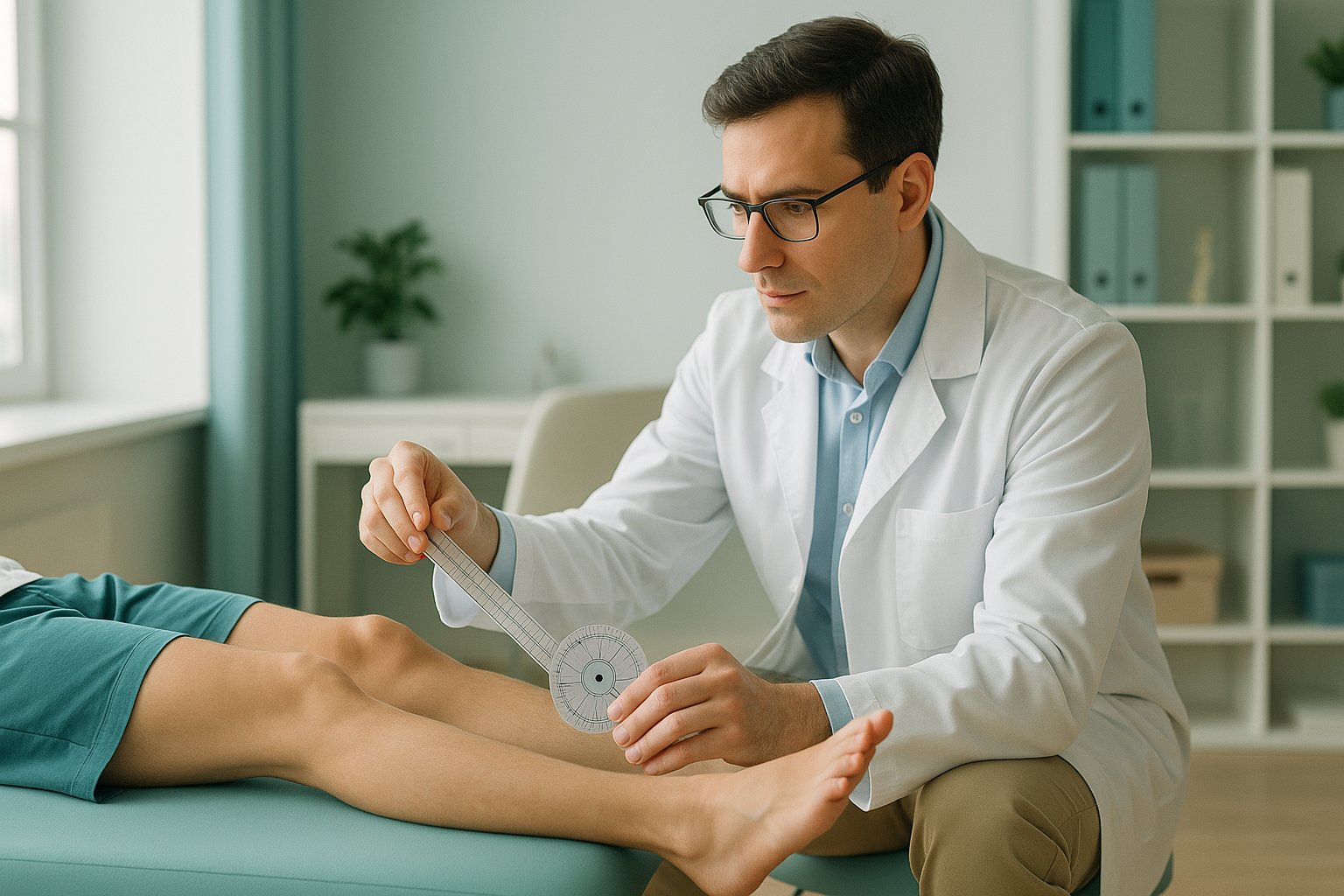
What measurements does an orthotist alignment report include?
The report includes true bone lengths for each femur and tibia, pelvic obliquity, the mechanical axis of each lower limb, and the overall sagittal and coronal balance of the spine. Every measurement comes from calibrated, full-length standing images. So the values reflect how your patient's skeleton actually behaves under gravity.
Femur and tibia measured separately, left against right, from calibrated standing images.
How much the pelvis tilts side to side under load, which directly affects shoe raise and AFO decisions.
The load line through hip, knee and ankle for each limb, essential for knee brace and prosthetic alignment.
Coronal and sagittal balance, showing how the spine compensates for lower-limb asymmetry.
All values come from one standing capture, so every measurement is internally consistent. There is no stitching of separate films and no magnification error from different source-to-film distances.
How do orthotists and prosthetists apply this data clinically?
The data feeds directly into prescription decisions where accuracy matters most. Below are the situations where standing alignment imaging adds the most value to your clinical workflow.
A verified bone length difference helps you choose the correct raise height, rather than relying on block tests alone.
Mechanical axis data confirms the correction angle your brace needs to achieve, improving comfort and function.
For transtibial and transfemoral prostheses, standing images show how socket position affects the overall load line.
A follow-up scan with the device in situ shows whether correction targets have been met.
Can you compare before and after scans to verify an intervention?
Yes. EOS imaging is calibrated and standardised. So a follow-up scan taken with the orthosis, prosthesis or shoe raise in place can be compared directly with the baseline. You can see, in exact terms, whether the pelvis has levelled, the mechanical axis has shifted and the spine has responded.
This before and after comparison is useful for audit, for shared care discussions with the referring consultant, and for explaining the effect of your intervention to the patient in a way they can clearly understand.
Referring clinicians: If you would like to set up a referral pathway for your patients, you can register to refer patients through our secure portal.
How do I refer a patient for a standing alignment scan?
You can refer a patient directly. No referral from the patient's doctor is needed. The process is simple.
Request the scan
Send a referral through the portal, or contact the clinic with your clinical question and the body regions you need imaged.
Patient attends
Your patient comes to ScanAlign, full-body standing imaging at 19 Harley Street, London. The scan takes under a minute and uses about 90% less radiation than a standard X-ray, around ten times less.
Report delivered
You receive a detailed alignment report with all the measurements relevant to your prescription, set out for clinical use.
Follow-up scan (optional)
After fitting the device, a repeat scan lets you verify the correction and record the outcome objectively.
A note on soft tissue. EOS imaging shows bones, joints and alignment. If you also need to assess soft tissue, discs or nerves, MRI is usually the better tool. The two work together, and both can be arranged at the same location.
Before your patient starts treatment, or changes the one they are on, a detailed diagnosis is worth having. Knowing whether the cause sits in the pelvis, the legs or the spine changes what will actually help.
Frequently asked questions
An EOS scan captures the full skeleton in one standing image, from head to feet. A conventional X-ray images one area at a time, and the patient may be lying down. EOS instead shows the whole alignment chain under real weight-bearing load. It also uses about 90% less radiation than a standard X-ray, around ten times less.
No. As a registered healthcare professional, you can refer directly. Patients can also refer themselves. No letter from their doctor is required.
EOS provides calibrated, full-length images with millimetre-level measurement accuracy. The whole limb is captured in one exposure, so there is no magnification error or stitching artefact. Both of these can affect measurements taken from conventional films.
Yes. The patient can stand in the scanner wearing their device. This is how before and after comparison works. The baseline scan is taken without the device, and the follow-up is taken with it in situ, so you can see the actual effect on alignment.
EOS uses about 90% less radiation than a standard X-ray, around ten times less. This low dose makes it well suited to follow-up imaging when you need to verify an intervention without unnecessary exposure.
The report is a structured clinical document with annotated images and numerical measurements. It is designed to be directly useful for prescription and shared care discussions.
The actual exposure takes under a minute. Most patients are in and out of the scanner room within a few minutes. The patient needs to stand still during the scan, and no special preparation is required.
The price for patients travelling from abroad is confirmed during the free video consultation, and you can find more on the ScanAlign FAQs and cost page.
They serve different purposes. EOS excels at showing bony alignment, joint geometry and mechanical axes under load. MRI is the better choice for soft tissue, disc pathology and nerve involvement. Many patients benefit from both.
ScanAlign is at 19 Harley Street, London. The clinic operates under The Harley Street Hospital's CQC licence.
Ready to refer a patient?
Set up a referral pathway or discuss a clinical case with our team. No obligation.
Book a free video consultationSources
- Illés, T. & Somoskeöy, S. "The EOS imaging system and its uses in daily orthopaedic practice." International Orthopaedics (SICOT), 2012; 36(7): 1405-1411.
- Sabharwal, S. & Kumar, A. "Methods for assessing leg length discrepancy." Clinical Orthopaedics and Related Research, 2008; 466(12): 2910-2922.
- International Society for Prosthetics and Orthotics (ISPO). "Standards for lower-limb prosthetic alignment." ispoint.org.
Written by Abbas Dhami (Specialist Diagnostic Radiographer)
This page is general information and not a diagnosis.
ScanAlign is the trading name of CoreMed Solutions Ltd and operates under The Harley Street Hospital's CQC licence.