
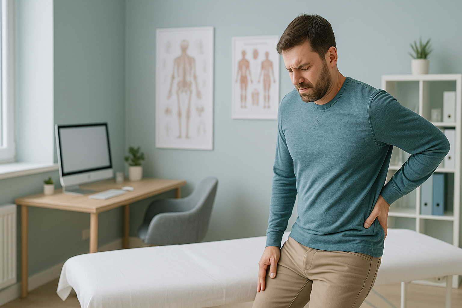
Why Does My Pain Change Depending on How I Stand or Sit?
Your pain changes with position because each posture moves the load onto different parts of your spine. Standing puts more pressure on the facet joints and narrows the spinal canal. Sitting puts more pressure on the discs. When your alignment is uneven, these shifts in load become more extreme. Standard scans taken lying down can miss them completely.
Key points
- Pain that changes with position is real, not imagined. Gravity and posture change how load spreads through your spine.
- Pain that is worse when standing often points to the facet joints or a narrowed spinal canal. Pain that is worse when sitting often points to pressure on the discs.
- Scans taken lying down remove gravity, so the problem may not show up on the images.
- Weight-bearing imaging shows your spine under real, everyday load. It reveals alignment problems that explain symptoms linked to position.
- Knowing your own pattern of which way makes it better or worse is the first step to the right diagnosis and the right care.
What is position-dependent pain?
Position-dependent pain is discomfort that clearly gets better or worse when you change posture. You might feel fine sitting but dread standing in a queue. Or your back may feel great on a walk but seize up the moment you sit at your desk.
This is not random. Your spine is a stack of bones, discs and joints held together by muscles and ligaments. When you change position, gravity acts on that stack in a different way. Some parts get squeezed. Others get stretched. If one part of the chain is worn, stiff or out of line, the load gathers there. That is where you feel it.

Clinicians sometimes call this a "directional preference". It is a truly useful clue. If a physiotherapist or surgeon knows which positions make you worse, they can narrow down which part of the spine is likely causing the problem.
What are the signs that your pain is position-dependent?
The main sign is a clear pattern that repeats. Your symptoms follow the same rules each day, even if how strong they feel varies.
Aching in the lower back or legs that builds the longer you stand, then eases when you sit or bend forward.
Deep pain in the middle of the back, or pain travelling down a leg, that builds while sitting and eases when you stand or walk.
Discomfort that seems to shift from your lower back to your hip or down a leg, depending on what you are doing.
One posture, such as lying on your side with your knees bent, that reliably takes the pain away almost completely.
What causes pain to change with position?
Several parts of the spine are sensitive to load. The position you take decides which one carries the most force.
Sitting bends the spine forward and squeezes the front of each disc. If a disc is worn or bulging, this extra pressure can push on nearby nerves.
Standing and bending the spine backwards presses on the small facet joints at the back of each vertebra. Worn facet joints protest under this load.
When you stand, the spinal canal narrows slightly. If it is already tight from spinal stenosis, standing can squeeze the nerves.
If your overall sagittal balance is off, your muscles work overtime to keep you upright. This causes tiredness and pain in positions that take more effort.
Your body works as one connected chain from your feet to your head. A stiff hip can shift load onto the lower spine. A flattened curve in the lower back can overload the discs. The pain often shows up in a completely different place from the real cause. This is why imaging of a single area, taken lying down, can tell only part of the story.
How does standing imaging reveal what lying-down scans miss?
Standing imaging captures your skeleton under the real pull of gravity. These are the same forces acting on you when your symptoms are at their worst. Imaging taken lying down removes gravity. Your muscles relax, your discs decompress and your joints separate slightly. The very alignment problems behind your positional pain can disappear from the picture.
An EOS scan images your full body from head to foot while you stand naturally. Gravity pulls on every joint, so any tilt, twist, shift or narrowing can be seen. Your clinician sees exactly how your spine and pelvis behave under the loads that trigger your symptoms.
The scan uses about 90% less radiation than a standard X-ray, around ten times less. It takes less than a minute and needs no preparation.
A standard X-ray, CT or MRI is taken lying flat. Without gravity, your spine settles into a resting position. A disc bulge may shrink. A narrowed canal may open up. Alignment shifts that only appear under load are not recorded. This is a key difference in weight-bearing versus lying-down imaging.
If your pain clearly changes with position, a weight-bearing scan shows your body in the posture that matters most.
Book a free video consultationWhat helps with position-dependent pain?
The first step is understanding the pattern. Once you know which parts are under stress, and in which positions, the right professional can build a plan around that. ScanAlign does not provide treatment. But a clear, whole-body standing image gives your physiotherapist, osteopath or surgeon the map they need.
- Find your pattern. Keep a simple note of which positions make your pain better or worse, and how long you can stay in each before symptoms build.
- Get the full picture. A whole-body standing image from ScanAlign, weight-bearing standing imaging captures your alignment under gravity, so nothing is hidden.
- Share it with your clinician. The report and images come straight to you. You can share them with any clinician you choose, wherever you are treated.
When MRI is the better tool. EOS shows bones, joints and alignment under load. If your clinician suspects a soft-tissue cause, such as a disc pressing on a nerve, a torn ligament or inflammation, MRI is usually the better test. The two work together. They do not compete.
Before you start a treatment, or change the one you are on, it is worth getting a detailed diagnosis first. Knowing whether the cause sits in your pelvis, your legs or your spine changes what will actually help.
Frequently asked questions
Standing bends the spine slightly backwards. This presses on the facet joints and narrows the spinal canal. If either is already worn or tight, standing adds load to it and the pain builds. Sitting bends the spine forward and takes pressure off those parts, so the pain eases.
Sitting raises the pressure inside the spinal discs because your lower back bends forward. Walking gently straightens the spine and spreads load more evenly across the discs and facet joints. This often reduces symptoms that come from the discs.
MRI is excellent for soft tissue, but it is taken lying down. With gravity removed, alignment shifts and canal narrowing that happen only under load may not appear. A weight-bearing scan captures the spine in the position that triggers your symptoms.
Directional preference means your pain keeps getting better with movement in one direction, such as bending backwards, and worse in the opposite direction. It is a real clinical sign that helps show which part of the spine is under stress.
In most cases, positional pain points to a mechanical cause, such as facet wear, disc changes or uneven alignment. But if you have sudden numbness, weakness, or loss of bladder or bowel control, get urgent medical help from your doctor or the nearest emergency department.
You stand still inside the scanner while it captures a full-body image from head to foot. It takes less than a minute, uses about 90% less radiation than a standard X-ray, and needs no special preparation.
No. ScanAlign accepts self-referrals. You can book a weight-bearing scan directly and share the results with any clinician you choose.
The price for patients travelling from abroad is confirmed during the free video consultation. Your first step is that free video consultation, where the team can confirm whether a scan is right for you.
Your body is a connected chain. When you change position, load shifts from one part to another. A compressed nerve in the lower spine may cause pain that spreads into the leg. How much the nerve is compressed can change depending on whether you are standing, sitting or bending.
EOS uses about 90% less radiation than a standard X-ray, around ten times less. This makes it one of the lowest-dose options available for imaging the skeleton. Your radiographer will discuss your history during the free video consultation.
See your spine the way it works
If your pain changes with position, a standing whole-body scan shows exactly what is happening under load. Start with a free video consultation from home before you plan your trip to London.
Book a free video consultationWritten by Abbas Dhami (Specialist Diagnostic Radiographer)
Sources
- Wilke, H-J et al. "New in vivo measurements of pressures in the intervertebral disc in daily life." Spine, 1999; 24(8): 755–762.
- National Institute for Health and Care Excellence (NICE). Low back pain and sciatica in over 16s: assessment and management (NG59), updated 2020.
- Illés, T & Somoskeöy, S. "The EOS imaging system and its uses in daily orthopaedic practice." International Orthopaedics, 2012; 36(7): 1405–1411.
This page is general information and not a diagnosis.
ScanAlign is the trading name of CoreMed Solutions Ltd and operates under The Harley Street Hospital's CQC licence.