
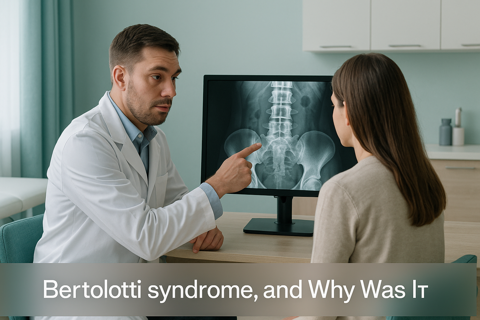
What Is Bertolotti Syndrome, and Why Was It Missed?
Bertolotti syndrome is lower back pain caused by a lumbosacral transitional vertebra. This is where the lowest lumbar bone is partly or fully fused to the sacrum. It is often missed because standard scans only show a small area. This makes it easy to miscount the vertebrae and miss the abnormal joint completely.
Key points
- A transitional vertebra is a normal variation in anatomy. It is found in roughly 4 to 35 per cent of people, but it does not always cause pain.
- When it does cause pain, the condition is called Bertolotti syndrome.
- The most common reason it is missed is an error in counting the vertebrae on a limited scan.
- A full spine x-ray taken while you stand lets the reporting clinician count every vertebra from top to bottom and see your alignment under load.
- An accurate diagnosis changes the treatment plan, including which level of the spine is targeted for any procedure.
What is a lumbosacral transitional vertebra?
A lumbosacral transitional vertebra (LSTV) is a bone at the very bottom of the spine. It does not look quite like a normal lumbar vertebra or a normal sacral segment. Instead, it sits somewhere in between. The lowest lumbar vertebra (usually called L5) may have an enlarged side process. This forms a partial or complete bony bridge to the sacrum below. This is called sacralisation of L5. In other cases, the top segment of the sacrum separates and behaves more like a lumbar vertebra. This is known as lumbarisation of S1.
Both are common. Many people have one and never know, because it causes no symptoms at all. When an LSTV does cause pain, usually on one side of the lower back, that is Bertolotti syndrome.

What does Bertolotti syndrome feel like?
The main sign is lower back pain on one side, often just above the belt line. It starts in the late teenage years or early adulthood. It tends to get worse with activity and ease with rest. The abnormal joint (sometimes called a pseudoarthrosis) can irritate nearby structures, so pain may also spread into the buttock or leg.
Usually felt to the left or right of the spine, not right in the middle. Often worse on the side of the bony bridge.
Bending, twisting or standing for a long time can make it worse. Sitting or lying down often brings relief.
The abnormal joint may irritate the nearby nerve root or sacroiliac area, sending pain into the buttock or thigh.
Symptoms often start in the late teens or twenties. This can lead clinicians to dismiss them as a muscle strain.
These symptoms overlap with disc problems and SI joint dysfunction. So Bertolotti syndrome is rarely the first thing considered.
Important. If you have sudden numbness, weakness in your legs, or any loss of bladder or bowel control, do not wait for a scan. Contact your doctor, call your local emergency number, or go to the nearest emergency department straight away.
Why is Bertolotti syndrome so often missed?
The biggest single reason is an error in counting the vertebrae. When you have a transitional vertebra, the normal numbering system breaks down. Most standard lumbar x-rays and MRI scans only show the lower part of the spine. Without seeing the full picture from top to bottom, the reporting clinician cannot reliably count down from C1. They might label your transitional L5 as a normal sacral segment, or label S1 as L5. That error of one level can mean the wrong disc, the wrong joint or the wrong nerve root is named as the problem.
An x-ray or MRI of the lower back alone cannot count vertebrae from the top of the spine. So the transitional bone may be labelled wrongly.
When you lie flat, weight is taken off the abnormal joint. The stress across the pseudoarthrosis disappears, and alignment can look normal.
The pain pattern looks like disc bulges, facet joint problems and SI joint pain. So scans are read with those diagnoses in mind.
Research suggests that numbering errors happen in a significant proportion of patients with a transitional vertebra when only a limited lumbar scan is done. This matters most if an injection or surgery is planned. Targeting the wrong level can mean the procedure fails, or a normal structure is treated without need.
How does a standing full spine scan help diagnose Bertolotti syndrome?
A standing full spine image captures every vertebra, from the base of the skull to the pelvis, in a single exposure while you carry your full body weight. This gives the reporting clinician two things that limited scans cannot.
Accurate counting of the vertebrae. With every vertebra visible, the clinician counts from C1 at the top all the way down. A transitional vertebra becomes obvious, rather than hidden at the edge of a cropped image.
Alignment under body weight. The body is a connected chain. A bony bridge on one side of L5 can tilt the pelvis, shift the spine sideways and force other areas to compensate. These shifts only appear when you are standing. Lying down relaxes the muscles and removes gravity. This often hides the very imbalance that is driving your pain.
At ScanAlign, unstitched full-body standing imaging uses EOS technology to capture this full view with about 90% less radiation than a standard x-ray, around ten times less. The image is a single, unstitched exposure, not several smaller x-rays joined together. This means no overlap errors and reliable measurement from head to pelvis.
If you have unexplained lower back pain and want a clear picture of your whole spine under load, a standing scan is a sensible first step.
Book a free video consultationWhen MRI is the better choice. EOS shows bones, joints and alignment clearly. If your clinician suspects a disc bulge, nerve compression or soft-tissue inflammation along with a transitional vertebra, MRI is usually the right tool for that part of the picture. The two scans work well together.
What can be done once Bertolotti syndrome is confirmed?
The first and most important step is an accurate diagnosis. ScanAlign is a diagnostic imaging clinic, not a treatment provider. The role of the standing scan is to give you and your clinician a precise map of your anatomy. Once the transitional vertebra is confirmed and correctly numbered, your treatment team can make informed decisions.
Common treatment options, guided by your specialist, may include physiotherapy to strengthen the surrounding muscles, guided injections at the correct joint to confirm the source of pain, or, less commonly, surgery at the correctly identified level. The key point is that every one of these options depends on knowing exactly which bone and which joint is responsible. A numbering error can send treatment in the wrong direction completely.
If you have been told your scans are normal but you still have persistent one-sided back pain, it may be worth asking whether the full spine was visible and whether a transitional vertebra was considered. You can book a full spine standing scan without a referral from your doctor.
Before you start treatment, or change the one you are on, it is worth getting a detailed diagnosis. Knowing whether the cause sits in the pelvis, the legs or the spine changes what will actually help.
Frequently asked questions about Bertolotti syndrome
Not exactly. A lumbosacral transitional vertebra is the variation in anatomy. Bertolotti syndrome is the diagnosis given when that variation is actually causing pain. Many people have a transitional vertebra without any symptoms.
Sacralisation of L5 means the lowest lumbar vertebra has partly or fully fused to the sacrum below. The side process of L5 is enlarged and forms a bony connection. It is one form of lumbosacral transitional vertebra.
Lumbarisation of S1 is the opposite pattern. The top segment of the sacrum partly separates and takes on the look and movement of a lumbar vertebra. This gives the impression of an extra lumbar bone.
Spinal procedures target a specific level. If the vertebrae are miscounted because of a transitional bone, an injection or surgery could be done at the wrong level. A full spine image removes this risk by letting the clinician count from the top down.
An MRI can show an enlarged transverse process and disc changes. But if the field of view is limited to the lumbar spine, the level may be miscounted. A full spine standing image is the most reliable way to confirm the numbering.
No. ScanAlign accepts self-referrals. You start with a free video consultation to check that the scan is suitable for your situation.
The EOS system uses about 90% less radiation than a standard x-ray, around ten times less. This makes it suitable for full spine imaging with a very low dose.
A transitional vertebra is present in a significant number of people. Among young adults with unexplained lower back pain, it is an under-recognised cause that should always be considered.
The price for patients travelling from abroad is confirmed during your free video consultation. You can also read the ScanAlign FAQs page.
Yes. When the transitional vertebra fuses on one side, it changes how load passes through the pelvis. This can put extra stress on the sacroiliac joint on the opposite side, sometimes causing SI joint dysfunction along with the syndrome.
Still wondering if your pain has been properly investigated?
A free video consultation takes just a few minutes, before you plan any trip to London. We will review your situation and tell you whether a standing full spine scan could help.
Book a free video consultationWritten by Abbas Dhami (Specialist Diagnostic Radiographer)
Sources
- Konin GP, Walz DM. Lumbosacral transitional vertebrae: classification, imaging findings, and clinical relevance. American Journal of Neuroradiology (AJNR). 2010;31(10):1778-1786.
- Castellvi AE, Goldstein LA, Chan DPK. Lumbosacral transitional vertebrae and their relationship with lumbar extradural defects. Spine. 1984;9(5):493-495.
- NHS. Low back pain in adults: assessment and management. NICE guideline NG59. National Institute for Health and Care Excellence, 2020 (updated).
This page is general information and not a diagnosis.
ScanAlign is the trading name of CoreMed Solutions Ltd and operates under The Harley Street Hospital's CQC licence.