
How Is a Child's Hip Monitored in Cerebral Palsy?
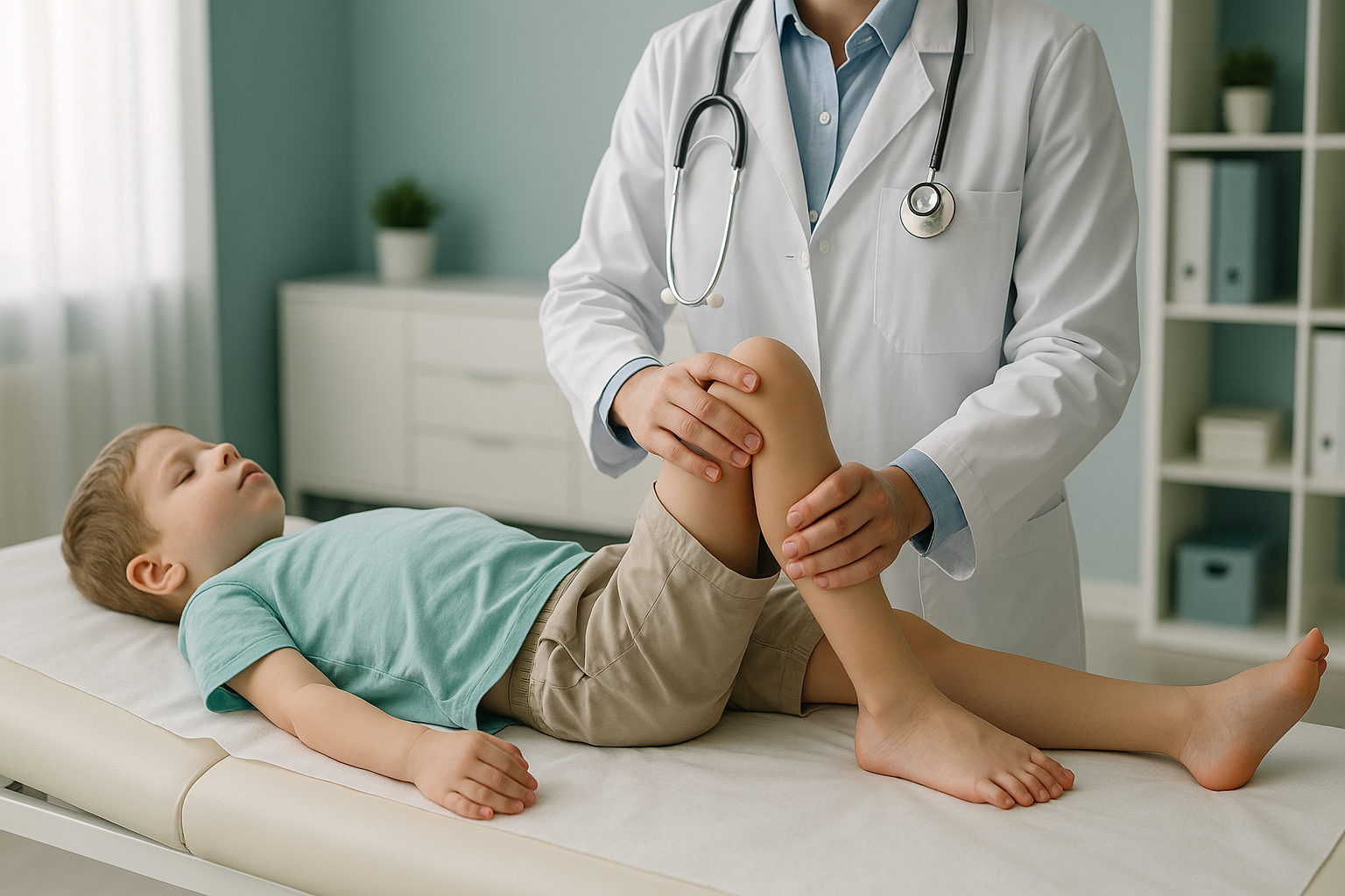
In cerebral palsy, hip surveillance uses regular X-rays to check whether the hip is slowly moving out of its socket. A measurement called the migration percentage tells the specialist team how far the hip has shifted. Finding this early gives the team time to act before pain or joint damage develops.
Key points
- Children with cerebral palsy have a high risk of hip displacement, especially at higher GMFCS levels.
- Hip surveillance is a planned programme of repeat X-rays. It usually starts in early childhood.
- The migration percentage is the main measure used to follow the hip's position over time.
- EOS imaging uses about 90% less radiation than a standard X-ray, around ten times less. This matters when scans are repeated over years.
- ScanAlign can scan children who walk and can stand for a short time. Children who cannot walk usually need lying-down (supine) imaging at their specialist centre.
What is hip surveillance in cerebral palsy?
Hip surveillance is a planned programme of regular clinical examinations and X-rays. Its aim is to find hip displacement early. In cerebral palsy, the muscles around the hip joint can pull unevenly because of differences in tone and spasticity. Over time, this imbalance can slowly draw the ball of the thighbone out of its socket.
The programme is led by your child's specialist team. This usually includes a paediatric orthopaedic surgeon, a physiotherapist and a paediatrician. X-rays are taken at set intervals and compared over time. This way, even very slow changes are noticed before they cause pain, stiffness or loss of function.
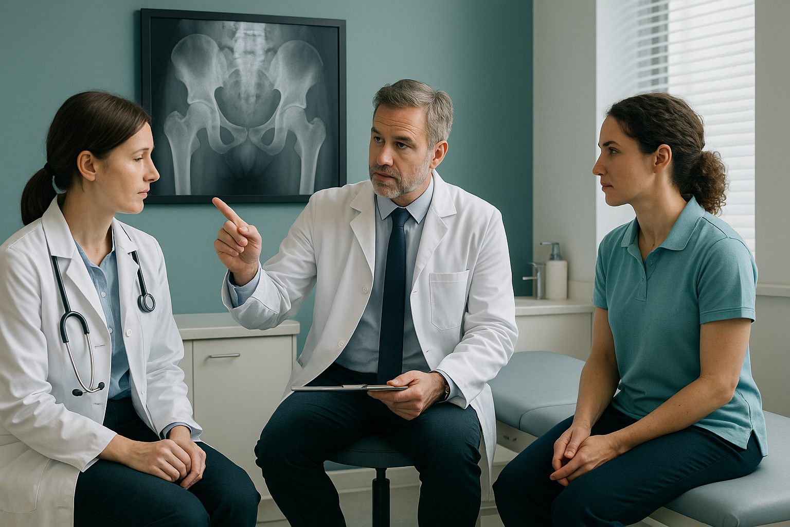
Hip displacement in cerebral palsy is not usually caused by an injury. It happens slowly, which is why ongoing surveillance is so important. Other hip conditions in children, such as developmental dysplasia of the hip, also benefit from early detection, although the underlying causes are different.
Why does the GMFCS level affect the risk of hip displacement?
The Gross Motor Function Classification System (GMFCS) grades how independently a child can move. It runs from Level I (walks without restrictions) to Level V (transported in a wheelchair). Children at higher GMFCS levels have more muscle imbalance around the hip and spend less time bearing weight. This raises the chance of hip displacement.
Walks independently. Lower risk, but surveillance is still recommended.
Walks with a hand-held aid. Moderate risk of displacement.
Uses a wheelchair. Highest risk. More frequent imaging recommended.
Research shows that up to 90% of children at GMFCS Level V develop some degree of hip displacement. Even children who walk independently can be affected. That is why surveillance covers all GMFCS levels, only at different intervals.
What are the signs of hip displacement in cerebral palsy?
Hip displacement often starts without clear symptoms. By the time pain appears, the hip may already have shifted a lot. That is why imaging is used, rather than waiting for symptoms alone.
Discomfort during nappy changes, while being moved or lifted, or when sitting, that cannot be explained by other causes.
Difficulty spreading the legs apart, or legs that feel stiffer on one side.
Trouble sitting evenly, or a habit of leaning to one side in the wheelchair or chair.
One leg looks shorter than the other when the child is lying down.
Children with cerebral palsy may also develop scoliosis (a sideways curve in the spine). This can interact with the position of the pelvis and the alignment of the hips. The specialist team monitors both together.
How is hip displacement measured on an X-ray?
The main measurement is the migration percentage. It shows how much of the ball of the thighbone sits outside the edge of the hip socket. A higher percentage means more displacement. The specialist team follows this number from scan to scan and looks for any rising trend.
The child is placed in the same standard position for each X-ray, so each image can be fairly compared with the last one. Keeping the position the same matters. Even a small change in how the child lies or sits can affect the measurement.
If the migration percentage rises past a certain level, the team may discuss further options. These could include changes to physiotherapy, seating or splints, or, in some cases, surgery. ScanAlign does not provide treatment, but the images can form part of the information the specialist team needs.
Thinking about your child's next monitoring scan? Our team can talk with you about whether children's imaging at ScanAlign might suit your child's needs.
Book a free video consultationWhy does radiation dose matter when scans are repeated over years?
Hip surveillance involves regular X-rays, often every six to twelve months over many years. So the total (cumulative) radiation dose adds up. Lowering the dose for each scan matters. EOS imaging uses about 90% less radiation than a standard X-ray, around ten times less. Over a whole childhood of monitoring, that difference becomes significant.
Can every child be scanned standing up?
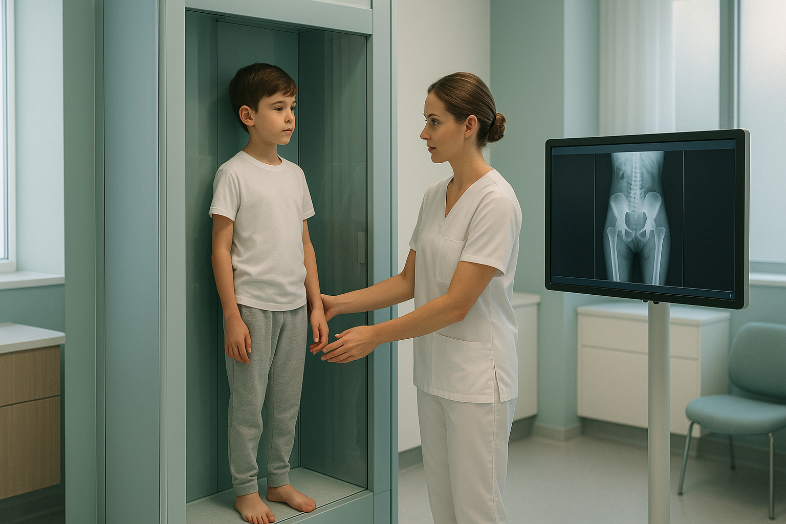
EOS takes images while the person stands upright. This shows how the joints and spine line up under the body's own weight. For children at GMFCS Levels I, II and sometimes III, who can stand for a short time with support, this can give useful information.
However, many children with cerebral palsy cannot stand. Children at GMFCS Levels IV and V, and some at Level III, usually need supine (lying-down) imaging. This is usually done at the child's specialist hospital. ScanAlign, low dose standing imaging, is best suited to children who walk or who can stand with support. During your free video consultation, we will tell you honestly whether our setup is right for your child, before you plan any trip to London.
When is MRI more suitable? EOS shows bones, joints and alignment clearly. For soft tissue, muscles, nerves or the spinal cord, MRI is usually the better tool. Your child's specialist team will decide which imaging is needed at each stage.
What the scan is like for a child
The scan itself takes only a few seconds. A parent or carer can stay in the room the whole time. The machine is open, not a tunnel, so it tends to be less stressful than other types of imaging. There is no noise, and your child does not need to keep still for a long time.
Before starting a treatment for your child, or changing the one they are on, it is worth getting a detailed diagnosis. Knowing whether the cause sits in the pelvis, the legs or the spine changes what will actually help.
Frequently asked questions
It is a planned programme of regular X-rays and clinical checks. It tracks whether the hip is slowly moving out of its socket. The aim is to catch any change early, before pain or joint damage happens.
The migration percentage shows how much of the ball of the thighbone sits outside the hip socket on an X-ray. A percentage that rises over time suggests the hip is slowly moving out of place.
It depends on your child's GMFCS level and earlier results. It is usually every six to twelve months, but your specialist team sets the schedule.
Not always, especially in the early stages. Many children have significant displacement before any pain is noticed. That is exactly why regular imaging is important.
Our EOS system takes images of people in an upright position. Children who can stand for a short time, even with support, may be suitable. Children who cannot stand at all usually need supine imaging at their specialist hospital. We talk about this openly in the free video consultation.
EOS uses about 90% less radiation than a standard X-ray, around ten times less. For children who need repeat imaging over many years, this helps keep the cumulative dose low.
No. You can book directly yourself. However, hip surveillance is usually managed by your child's specialist team, so we recommend discussing it with them. We are happy to share our images and reports with your team.
Yes. A parent or carer is welcome in the room for the whole scan. The system is open, not enclosed, and the scan takes only a few seconds.
It can. Children with cerebral palsy may develop scoliosis, a sideways curve in the spine. The specialist team often monitors the spine and hips together because they affect each other.
For patients travelling from abroad, the price is confirmed during the free video consultation. You can also visit our book a monitoring scan page.
Not sure if ScanAlign is right for your child?
In our free video consultation, you speak with a specialist radiographer. They will talk honestly about your child's needs, including whether standing imaging is suitable, so you can decide before travelling to London.
Book a free video consultationWritten by Abbas Dhami (Specialist Diagnostic Radiographer)
Sources
- Wynter M, et al. "Australian hip surveillance guidelines for children with cerebral palsy." Journal of Pediatric Rehabilitation Medicine, 2014.
- Hägglund G, et al. "Prevention of dislocation of the hip in children with cerebral palsy: 20-year results of a population-based prevention programme." The Bone & Joint Journal, 2005.
- National Institute for Health and Care Excellence (NICE). Cerebral palsy in under 25s: assessment and management (NG62), 2017.
This page is general information and not a diagnosis.
ScanAlign is the trading name of CoreMed Solutions Ltd and operates under The Harley Street Hospital's CQC licence.