
What Is Cervical Instability, and How Is It Assessed?
Cervical instability means there is more movement than normal between two or more vertebrae in your neck. It can cause neck pain, headaches and a feeling that your head is too heavy. The standard way to assess it is with flexion-extension X-rays. These capture the neck as you bend it forward and back, and show movement that ordinary imaging may miss.
Key points
- Cervical instability is too much movement between neck vertebrae. It is not simply a stiff or sore neck.
- Flexion-extension X-rays are the accepted way to check for abnormal movement.
- It is more common in people with generalised hypermobility syndrome or connective tissue conditions.
- Imaging taken lying down can miss instability, because gravity and muscle tone change when you stand.
- A referral to a specialist is usually needed for diagnosis and management.
Your cervical spine is the top seven vertebrae of your neck. They support the weight of your head. They let you look up, down and to either side. Between each pair of vertebrae, ligaments, small joints and discs keep everything in place. When these structures are damaged, stretched or naturally lax, the vertebrae can shift more than they should. That extra movement is cervical instability.
The term also covers two specific areas that specialists look at closely. Craniocervical instability describes extra movement where the skull sits on the top vertebra (C1). Atlantoaxial instability describes extra movement between C1 and C2, the two uppermost vertebrae. Both are assessed in the same way, with careful imaging and clinical examination.

What are the symptoms of cervical instability?
Symptoms vary. They depend on which level of the neck is affected and how much extra movement there is. Many people describe a group of problems rather than a single symptom.
A constant feeling that your head is too heavy for your neck, especially by the end of the day.
A deep ache in the upper neck, often worse after sitting upright or looking at a screen.
Feeling off-balance, light-headed or disoriented when you turn your head.
Persistent headaches that start at the back of the head and can spread forward.
Some people report blurred vision, trouble concentrating or a foggy feeling.
When to seek urgent help
If you develop sudden weakness or numbness in your arms or legs, difficulty walking, loss of bladder or bowel control, or severe worsening symptoms, contact your doctor or go to the nearest emergency department straight away. These could be signs of spinal cord compression, which needs immediate attention.
What causes cervical instability?
Cervical instability happens when the ligaments, joints or other structures that hold the neck vertebrae together cannot do their job properly. This can be the result of injury, wear over time, or an underlying condition that affects connective tissue.
Whiplash, falls or sports injuries can stretch or tear the ligaments that keep the upper neck stable.
Conditions such as hypermobile Ehlers-Danlos syndrome make ligaments throughout the body more lax, including in the neck.
Years of wear on the discs and joints can slowly reduce the support around individual vertebrae.
Rheumatoid arthritis, for example, can wear away the ligaments at C1-C2 over time.
If you already have generalised joint hypermobility syndrome, your neck ligaments may be naturally more elastic. This does not automatically mean you have cervical instability. But it does mean the possibility should be considered if you have persistent symptoms.
How is cervical instability properly assessed?
The accepted standard is a clinical examination by a specialist, combined with flexion-extension imaging. The specialist is usually a spinal surgeon or a consultant in musculoskeletal radiology or neuroradiology. They will examine your neck movements and nerve function before asking for the right imaging.
Flexion-extension X-rays
These are X-rays taken while you gently bend your neck forward (flexion) and backward (extension). The two images are then compared. If a vertebra slides further forward or backward than the accepted limit, that segment is considered unstable. This is the standard test because a single still image, taken in a neutral position, may look completely normal even when significant movement is present.
Why a standard X-ray or MRI may not be enough
A routine neck X-ray captures one position only. An MRI is taken lying down. This removes the load of your head and lets the muscles relax. Both can miss instability that only shows up when the neck moves under its own weight. That is why flexion-extension views matter so much. They test the neck while it is actually doing its job.
A note on MRI. EOS and X-ray are excellent at showing bone position and alignment. For soft tissue, discs, the spinal cord or nerves, MRI is usually the better tool. Many people with suspected cervical instability will need both types of imaging at different stages of their assessment.
A word of caution about overdiagnosis
Craniocervical instability has received a lot of attention online, especially among people with hypermobility. It is a real and sometimes serious condition, but there is a risk of overdiagnosis. Not every headache or dizzy spell in someone with hypermobility means the upper neck is unstable. A proper assessment needs the right specialist, the right imaging and careful matching of the findings with your symptoms and examination. Be wary of any clinic that offers a diagnosis based on a single scan without a thorough clinical examination.
Can a standing scan help with neck and spine assessment?
Yes, but it is important to be clear about what a standing scan does and does not do. A standing whole-body EOS scan captures your entire spine, from skull to pelvis, while you are upright and bearing weight. This shows how your cervical lordosis (the natural curve of your neck) relates to the rest of your spinal alignment and posture under gravity.
When you lie down for a standard X-ray or MRI, gravity is removed and muscles relax. This can hide the way your spine actually behaves in daily life. A standing image shows how the body compensates. For example, if your lower back is unusually flat, your neck may curve more than expected to keep your head balanced. Seeing this whole chain gives useful context to any specialist assessing your neck.
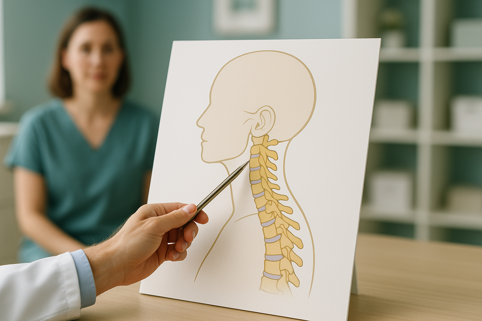
A standing EOS scan does not replace dedicated flexion-extension views. It does not test movement. What it does is give a detailed, low-dose standing image of your full spinal alignment, using about 90% less radiation than a standard X-ray, around ten times less. This wider picture can be shared with your specialist alongside other imaging, to build a fuller understanding of your neck problem.
If you would like to see how your full spine aligns while standing, you can book a standing neck and spine scan at our clinic at 19 Harley Street, London. Before you plan your trip, we will discuss your situation in a free video consultation.
Book a free video consultationWhat can help with cervical instability?
Management depends entirely on how severe the problem is and what is causing it. It is guided by a specialist. ScanAlign is a diagnostic imaging clinic and does not provide treatment. The information below is general only.
- Specialist referral. A spinal surgeon, neurologist or rheumatologist with experience of cervical instability should oversee your care.
- Physiotherapy. Strengthening the deep neck flexor muscles and improving postural control can give more support to the cervical spine. This should be supervised by a physiotherapist who understands the condition.
- Collar use. In some cases a soft or rigid collar may be recommended for short periods, but this is a specialist decision. Wearing a collar for a long time without guidance can weaken the muscles further.
- Further tests. Depending on the findings, upright or dynamic MRI, CT, or other specialist imaging may be needed.
- Surgery. In severe or progressive cases, a spinal surgeon may discuss fusion to stabilise the affected segment. This is kept for clearly confirmed instability with significant symptoms.
Before you start treatment, or change the one you are on, it is worth getting a detailed diagnosis. Knowing whether the cause sits in the pelvis, the legs or the spine changes what will actually help.
Frequently asked questions about cervical instability
Most people describe a heavy or wobbly feeling in the head, deep neck pain, headaches at the base of the skull, and sometimes dizziness or brain fog. Symptoms are often worse after being upright for a long time or after holding the head in one position.
Not quite. Cervical instability is a broader term that covers any level in the neck. Craniocervical instability describes extra movement where the skull meets C1. Atlantoaxial instability means extra movement between C1 and C2. All are assessed in a similar way.
Generalised hypermobility syndrome and connective tissue conditions like hypermobile Ehlers-Danlos syndrome can make ligaments throughout the body more lax, including those in the cervical spine. This does not mean everyone with hypermobility has cervical instability. But it raises the possibility if symptoms are present.
Flexion-extension X-rays are the accepted standard. They capture the neck bending forward and backward, and show whether a vertebra moves beyond normal limits. An MRI may also be needed to assess the spinal cord, discs and ligaments. A single still X-ray in a neutral position is not enough.
A standard MRI taken lying flat shows the spine without load. Instability often only shows up under load or during movement, so a lying-down MRI alone may not capture the extra movement. Even so, MRI is still essential for assessing soft tissue, the spinal cord and nerves.
An EOS scan is a single standing image. It does not test movement, so it cannot diagnose instability on its own. What it does is show your full spinal alignment under gravity, including the position and curve of your cervical spine. This can give useful context to a specialist who is already investigating your neck.
No. You can refer yourself. We offer a free video consultation first, to discuss your situation and make sure a standing EOS scan is right for you.
An EOS scan uses about 90% less radiation than a standard X-ray, around ten times less. This makes it one of the lowest-dose options for full-body skeletal imaging.
The price for patients travelling from abroad is confirmed during the free video consultation.
A diagnosis should come from a qualified specialist using proper imaging, not from a single scan or an online assessment. If you have been given this diagnosis, ask to see the flexion-extension views and discuss the findings with a spinal surgeon or neurologist. Mild instability is often managed conservatively with physiotherapy. Severe cases may need closer monitoring or input from a surgeon.
See how your whole spine aligns while standing
A free video consultation lets us discuss your symptoms, and whether a standing EOS scan would add useful information for your specialist, before you decide to travel to London.
Book a free video consultationLearn more about ScanAlign standing EOS imaging and how whole-body alignment assessment works.
Written by Abbas Dhami (Specialist Diagnostic Radiographer)
Sources
- White AA, Panjabi MM. Clinical Biomechanics of the Spine. 2nd ed. Philadelphia: Lippincott; 1990. (The foundational reference for cervical stability criteria and measurement thresholds.)
- British Society of Skeletal Radiologists. Guidelines on flexion-extension radiography of the cervical spine. Available at: bssr.org.uk
- NHS. Neck pain: causes, treatment and when to get help. Available at: nhs.uk/conditions/neck-pain/
This page is general information and not a diagnosis.
ScanAlign is the trading name of CoreMed Solutions Ltd and operates under The Harley Street Hospital's CQC licence.