

How Does Diastasis Recti Affect Your Posture?
Diastasis recti is a gap between the two sides of your abdominal muscles. It weakens the support of your trunk. This lets the pelvis tilt forward and the curve of the lower back increase. It changes your standing alignment from head to feet. It often causes lower back pain that seems to have nothing to do with the abdomen itself.
Key Takeaways
- Diastasis recti means the left and right halves of your rectus abdominis have separated along the linea alba, the connective tissue running down the midline.
- The loss of core support that follows often leads to anterior pelvic tilt and increased lumbar lordosis.
- Pain commonly appears in the lower back, hips or pelvis rather than the abdomen.
- A standing, weight-bearing image of your whole alignment can show compensations that lying-down scans miss.
- Graded loading exercises guided by a specialist physiotherapist are the first-line approach. Surgery is only considered after sustained rehabilitation.
What is diastasis recti?
Diastasis recti is a widening of the gap between the two strips of the rectus abdominis muscle, often called the "six-pack." These strips are joined by a band of fibrous tissue called the linea alba. It runs straight down from the breastbone to the pubic bone. When this tissue stretches and thins, it can no longer hold the two muscle halves close together.
It is most often linked with pregnancy. But it also happens in men and in women who have never been pregnant. Repeated heavy lifting, rapid weight gain or long-term rises in intra-abdominal pressure can all play a part.
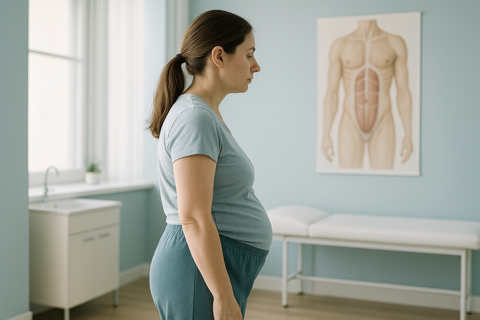
The gap is not a tear. The muscle itself is still intact, but the connective tissue between the two halves has become loose. This reduces the core's ability to create and pass on force, and that has a direct effect on your posture.
How do you check for diastasis recti?
For a simple self-check, lie on your back with your knees bent. Then gently lift your head and shoulders. Press your fingers along the midline above and below the navel. If you feel a gap wider than about two finger widths, or the tissue feels soft and gives little support, you may have diastasis recti. A physiotherapist or your doctor can confirm this with a clinical examination.
A ridge or dome along your midline when you brace or sit up from lying down.
Persistent aching across the lower back, especially after standing for a long time.
The pelvic floor and deep abdominal muscles work as a team. When one weakens, the other often follows.
Finding it hard to stand tall without thinking about it, or feeling that your trunk "sags" forward.
Why does diastasis recti cause back pain and posture changes?
Your abdominal muscles are a key part of the system that supports your trunk. They work with your back muscles, diaphragm and pelvic floor to keep your spine stable. When the front wall weakens because of diastasis recti, your body has to compensate.
The most common compensation is an increase in anterior pelvic tilt. Without enough abdominal bracing, the pelvis tips forward and the lower back curves more deeply. The upper back then rounds to balance. This chain of compensations can cause pain and stiffness in your lower back, hips, or between your shoulder blades, all far from the gap in your abdomen.
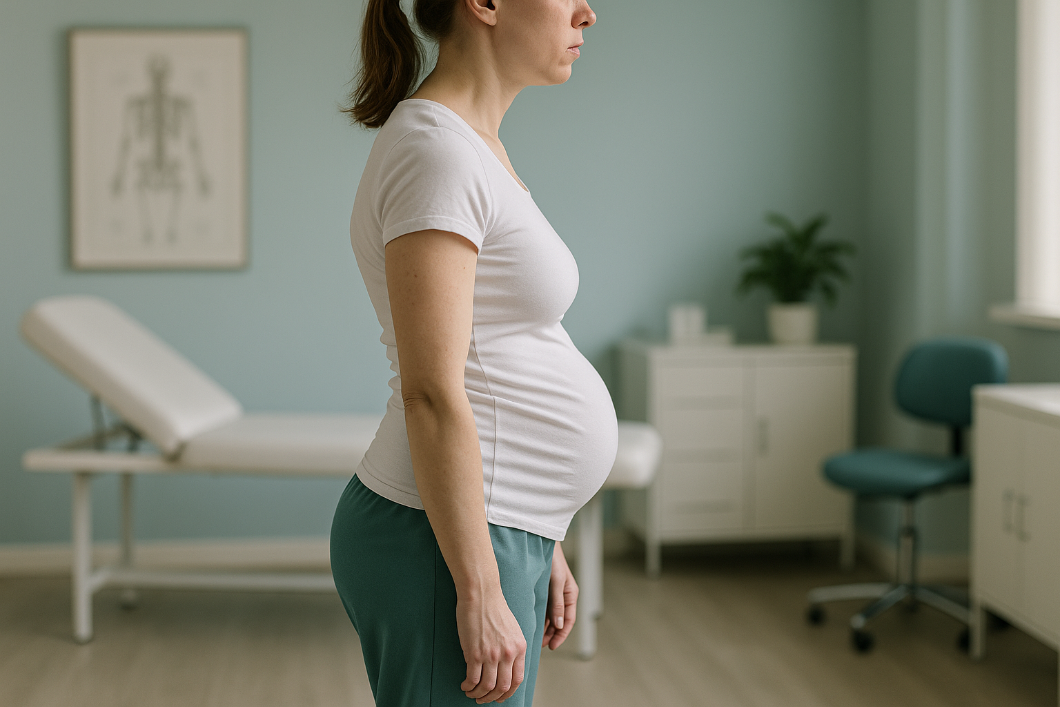
This is why lying-down scans can be misleading for posture problems. When you lie on a table, gravity flattens the spine, the muscles relax and the pelvic tilt disappears. The alignment problems that drive your symptoms only show up when you are standing and carrying your own weight.
What causes diastasis recti?
Anything that raises pressure inside the abdomen again and again, or for a long time, can stretch the linea alba. The best-known cause is pregnancy, but it is not the only one.
The growing uterus stretches the abdominal wall over many months. It is the most common cause of abdominal separation after birth.
Lifting heavy loads again and again without proper bracing technique can overload the midline tissue over time.
A quick increase in abdominal fat raises intra-abdominal pressure and stretches the connective tissue.
Persistent coughing, constipation or poor exercise form can all add to long-lasting abdominal pressure.
How is posture assessed when you have diastasis recti?
Diastasis recti changes your alignment while you are upright. So a lying-down scan tells only part of the story. A standing, weight-bearing image shows how your skeleton actually carries load under gravity. It shows pelvic tilt, changes in the lumbar curve and any knock-on effects up and down the chain.
ScanAlign standing EOS imaging takes a full-length image of your skeleton from head to feet while you stand naturally. It uses about 90% less radiation than a standard X-ray, around ten times less. The result is a detailed picture of your whole alignment, not just one area. Your clinician can then see where compensations are happening and plan rehabilitation to match.
Wondering whether your posture changes are linked to abdominal separation? A free video consultation from home can help you decide if travelling to London for a standing alignment assessment is the right next step.
Book a free video consultationWhen MRI is the better tool: EOS imaging shows bones, joints and alignment. If your clinician suspects soft-tissue problems such as disc bulges, nerve compression or muscle damage, MRI is usually the more suitable test. The two types of imaging often work well together.
What helps with diastasis recti and posture recovery?
The first-line approach is a programme of graded loading exercises. Ideally, it is guided by a physiotherapist who specialises in postnatal or abdominal rehabilitation. The aim is to retrain the deep core muscles to create tension across the linea alba, gradually closing the gap and restoring support to the trunk.
General principles of rehabilitation
- Graded loading. Exercises start gently and build up as the tissue adapts. Rushing into sit-ups or planks too early can make the gap worse.
- Breath and pelvic floor coordination. Learning to breathe and brace properly helps you manage intra-abdominal pressure during everyday movements.
- Posture awareness. Understanding how your pelvis, spine and ribcage relate to each other makes it easier to stand and move without too much compensation.
- Realistic timelines. Recovery takes months, not weeks. Progress is often measured by how well you function, not just by the width of the gap.
In persistent cases where rehabilitation has been fully tried, surgery may be considered. This decision is made with a specialist surgeon. It is not the starting point.
Before you start treatment, or change the one you are on, it is worth getting a detailed diagnosis. Knowing whether the cause sits in the pelvis, the legs or the spine changes what will actually help.
If your symptoms include sudden numbness, weakness in your legs, or loss of bladder or bowel control, get urgent medical help from your doctor or your nearest emergency department. These are not typical of diastasis recti alone and need prompt assessment.
Frequently asked questions about diastasis recti and posture
Yes. When the abdominal wall cannot support the spine properly, the lower back muscles work harder to make up for it. This commonly leads to persistent aching across the lower back, especially after standing or walking for long periods.
Lie on your back with your knees bent. Gently lift your head and shoulders and press your fingers along the midline above and below your navel. A gap wider than about two finger widths, or tissue that feels soft and gives way easily, may point to diastasis recti. A physiotherapist or your doctor can confirm this with an examination.
No. Pregnancy is the most common cause, but diastasis recti also happens in men and in women who have never been pregnant. Heavy lifting, rapid weight gain and long-term rises in abdominal pressure can all stretch the linea alba.
Traditional sit-ups, crunches and heavy front-loaded movements can increase pressure on the weakened midline. A specialist physiotherapist can assess your own gap and prescribe safe exercises that build up step by step. These strengthen the core without making the separation worse.
Some mild cases improve naturally within the first few months after birth as the tissue recovers. But wider separations, or those that last beyond several months, usually benefit from guided rehabilitation. Without targeted exercise, the gap and its effects on posture tend to remain.
The posture changes caused by diastasis recti, such as more pelvic tilt and a deeper lower back curve, only appear when you are standing and bearing weight. Lying-down imaging relaxes these compensations and can miss the full picture. A standing EOS scan captures your real-life alignment from head to feet.
Yes. The EOS system uses about 90% less radiation than a standard X-ray, around ten times less. It captures a full-length standing image in a single pass.
No. ScanAlign accepts self-referrals. You can book a postnatal alignment assessment directly through a free video consultation. A specialist radiographer will confirm whether a standing scan is right for you.
The price for patients travelling from abroad is confirmed during the free video consultation, before you commit to anything.
Yes. A weakened core lets the pelvis tilt and rotate unevenly. This can overload the hip joints and pelvic ligaments. Pain in the front of the hip, the SI joint or the groin is not uncommon when diastasis recti is present.
See how your whole body aligns, standing up
A free video consultation takes a few minutes, and you can join from home. A specialist radiographer will review your concerns and tell you whether a standing alignment scan could help, before you plan a trip to London.
Book a free video consultationSources
- Sperstad JB, Tennfjord MK, Hilde G, Ellström-Engh M, Bø K. Diastasis recti abdominis during pregnancy and 12 months after childbirth: prevalence, risk factors and report of lumbopelvic pain. British Journal of Sports Medicine. 2016;50(17):1092-1096.
- Benjamin DR, van de Water ATM, Peiris CL. Effects of exercise on diastasis of the rectus abdominis muscle in the antenatal and postnatal periods: a systematic review. Physiotherapy. 2014;100(1):1-8.
- NHS. Diastasis recti (abdominal muscle separation). NHS guidance on postnatal abdominal recovery. nhs.uk
Written by Abbas Dhami (Specialist Diagnostic Radiographer)
This page is general information and not a diagnosis.
ScanAlign is the trading name of CoreMed Solutions Ltd and operates under The Harley Street Hospital's CQC licence.