
What Is a Distal Femoral Osteotomy, and When Is It Chosen?
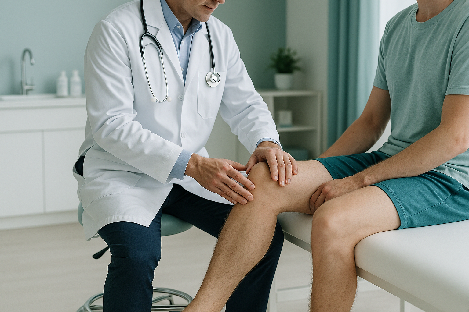
A distal femoral osteotomy is a knee operation that keeps your own joint. It reshapes the lower end of the thigh bone (femur) to correct valgus, or knock-knee, alignment. It moves the weight-bearing line back towards the centre of the knee. This reduces overload on the outer (lateral) compartment. In younger, active adults, it can delay or avoid a knee replacement.
Key takeaways
- A distal femoral osteotomy (DFO) corrects valgus knee alignment by reshaping the femur, not the tibia.
- It is a joint-preserving option for younger or more active adults who want to delay a knee replacement.
- Accurate standing alignment imaging is essential for planning before surgery.
- EOS imaging takes a full-leg standing x-ray while you stand naturally and bear your weight, with about 90% less radiation than a standard x-ray.
- ScanAlign does not perform surgery. It provides the diagnostic imaging that guides your surgical team.
What exactly is a distal femoral osteotomy?
A distal femoral osteotomy is a controlled surgical cut near the lower end of the femur, just above the knee joint. The surgeon removes or adds a small wedge of bone. Then a plate fixes the bone in its new position. This changes the angle of the thigh bone. The weight-bearing line of the leg then passes through the centre of the knee, instead of overloading the lateral (outer) compartment.
The word "distal" simply means "the end closest to the knee," and "osteotomy" means "bone cut." The operation keeps your own joint surfaces instead of replacing them. This is why it is often called joint-preserving knee surgery.
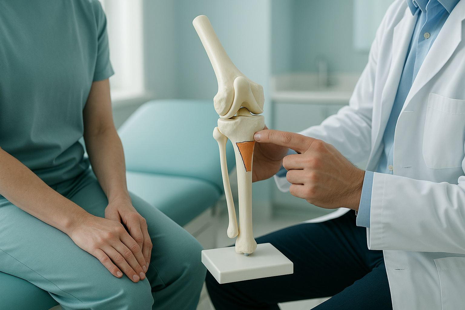
The goal is simple: spread the load across the knee so that healthy cartilage does more of the work and damaged cartilage is protected. For many younger adults, this buys valuable years of pain-free activity before a replacement becomes necessary.
How do you know your knee alignment might need correction?
Valgus alignment, commonly called knock knees, is not always obvious. Some people live with a mild angle for years before pain starts. The signs below may suggest that the outer compartment of your knee is under too much strain.
These signs do not confirm that you need a distal femoral osteotomy. They simply suggest that a proper alignment assessment, ideally while standing, could help find the structural cause.
What causes valgus knee alignment?
Valgus alignment develops when the weight-bearing line of the leg drifts to the outside of the knee centre. This can happen for several reasons.
Knowing the cause matters because it shows whether the problem sits in the femur, the tibia, or both. When the deformity starts in the femur, a distal femoral osteotomy deals with it at the source. If the tibia is the main contributor, a different procedure, such as knock knee surgery at the level of the tibia, may be more appropriate. Accurate imaging is the key to telling these apart.
What surgical techniques are used in a distal femoral osteotomy?
Surgeons usually choose between two methods. The choice depends on the pattern and size of the correction needed.
Opening wedge DFO. The surgeon makes a partial cut through the inner side of the femur, gently opens the gap, and fills it with bone graft or a substitute. A locking plate holds the new angle while the bone heals. This method allows fine adjustment and keeps the bone length.
Closing wedge DFO. A wedge of bone is removed from the outer side of the femur. The two surfaces are brought together and fixed with a plate. Healing can be faster because bone meets bone directly, but the leg may become very slightly shorter.
In both methods, the correction angle is worked out before surgery using precise standing images of the full leg. The surgeon traces the weight-bearing line, measures the current angle, and plans exactly how much correction is needed. This is why high-quality, weight-bearing imaging is so important.
Why does planning before surgery need standing imaging?
When you lie down for a usual x-ray or CT scan, gravity stops pulling on your joints. Your muscles relax and the leg may straighten slightly. This can hide the true valgus angle. A full-leg standing x-ray shows your alignment the way you actually use your legs, under your real body weight.
EOS imaging goes one step further. The scanner captures both legs from hip to ankle in a single upright exposure, with about 90% less radiation than a standard x-ray, around ten times less. Because the image is calibrated, surgeons can take accurate measurements directly from the scan without magnification errors.
At ScanAlign, standing EOS imaging in London, a specialist radiographer places you naturally in the scanner and produces a detailed report on your alignment from hip to foot. Your surgeon then uses this to plan the correction angle for a distal femoral osteotomy, or to decide whether a different approach suits you better.
Thinking about a femoral osteotomy? Accurate alignment imaging is the starting point.
Book a free video consultationWhen MRI may also be needed. EOS is ideal for bone alignment and joint spacing. If your surgeon needs to look at soft tissue, cartilage thickness, ligaments or nerves in detail, MRI is usually the better tool. The two scans complement each other.
Who is a good candidate for a distal femoral osteotomy?
A DFO is usually recommended for adults with valgus malalignment and damage mainly in the lateral compartment, who still have healthy cartilage on the inner side. It suits people who want to stay active and delay a partial or total knee replacement.
- Younger, active adults with valgus alignment
- Cartilage loss in the lateral compartment with a healthy medial side
- Patients who want to keep their own knee joint
- People whose deformity starts in the femur
- Advanced arthritis affecting both sides of the knee
- Significant stiffness or limited range of motion
- Smokers or people with poor bone healing potential (relative factor)
- Deformity that starts in the tibia rather than the femur
Only your orthopaedic surgeon can decide if a DFO is right for you. Imaging gives your surgeon the objective alignment data needed to make that decision with confidence. If you would like to travel to London to book alignment imaging before surgery, a short free video consultation is the first step.
What does recovery from a distal femoral osteotomy look like?
Recovery is different for each person. It depends on the technique used, the size of the correction, and your general health. Broadly, most people follow a gradual path back to activity.
- Early weeks. Crutches and limited weight-bearing to protect the healing bone. Physiotherapy starts soon after surgery.
- Middle phase. A gradual return to weight-bearing as x-rays confirm the bone is healing. Swimming and cycling are often brought back before impact sports.
- Longer term. Full activity, including running and sport, may be possible once the bone has fully healed and the muscles have regained strength.
Your surgical team will give you a personal timeline. ScanAlign does not perform surgery or manage rehabilitation. Its role is to provide the standing alignment imaging that supports planning before surgery and, if your surgeon asks for them, follow-up comparison scans.
Before you start any treatment, or change the one you are on, it is worth getting a detailed diagnosis. Knowing whether the cause sits in the pelvis, the legs or the spine changes what will actually help.
Seek urgent help if needed. If you have sudden severe knee pain, numbness, weakness in the leg, or loss of bladder or bowel control, contact your doctor, call your local emergency number, or go to the nearest emergency department. These symptoms need immediate medical attention, not a scan booking.
Frequently asked questions about distal femoral osteotomy
A distal femoral osteotomy corrects alignment by reshaping the lower femur. It is used mainly for valgus (knock-knee) deformities. A high tibial osteotomy reshapes the upper tibia. It is used mainly for varus (bow-leg) deformities. The choice depends on where the malalignment starts, and standing imaging shows this.
It is a significant operation done under general anaesthetic. The bone is cut and repositioned, then fixed with a metal plate. However, it is classed as joint-preserving surgery, meaning it keeps your own knee joint intact.
Recovery times vary. Most people use crutches for several weeks and slowly return to full weight-bearing over a few months. A full return to sport may take longer. Your surgeon will give you a personal plan.
That is one of its main purposes. It moves load away from the damaged compartment. This can reduce pain and slow further cartilage wear, possibly delaying knee replacement by many years.
When you lie down, gravity is removed and your alignment can look different from when you stand. Standing imaging shows the true weight-bearing angle of the leg. Surgeons use this measurement to work out exactly how much correction is needed.
An EOS scan uses about 90% less radiation than a standard x-ray, around ten times less. This makes it a particularly low-dose option for full-leg imaging.
No. ScanAlign is a private, self-pay clinic. You can refer yourself without a letter from your doctor or consultant. The process starts with a free video consultation.
The price for patients travelling from abroad is confirmed during the free video consultation.
EOS is designed for bones, joints and alignment. For a detailed look at soft tissue, including cartilage, ligaments and nerves, MRI is usually the better tool. The two scans work well together.
No. EOS technology is available at a small number of clinics in the UK. ScanAlign is a private, self-pay clinic at 19 Harley Street, London, focused entirely on standing whole-body alignment imaging.
Understand your alignment before surgery
A standing EOS scan gives your surgeon the precise measurements needed to plan a distal femoral osteotomy. Start with a free video consultation.
Book a free video consultationSources
- Wylie JD, Jones DL, Hartwell MJ, et al. "Distal Femoral Osteotomy for the Valgus Knee: Medial Closing Wedge Versus Lateral Opening Wedge." Arthroscopy, 2016;32(10):2037–2047.
- British Orthopaedic Association. Patient information on osteotomy around the knee. boa.ac.uk
- Deschamps G, Chol C. "Fixed-bearing unicompartmental knee arthroplasty. Patients' selection and operative technique." Orthopaedics & Traumatology: Surgery & Research, 2011;97(6):648–661. (Comparative context for joint-preserving options.)
Written by Abbas Dhami (Specialist Diagnostic Radiographer)
This page is general information and not a diagnosis.
ScanAlign is the trading name of CoreMed Solutions Ltd and operates under The Harley Street Hospital's CQC licence.