
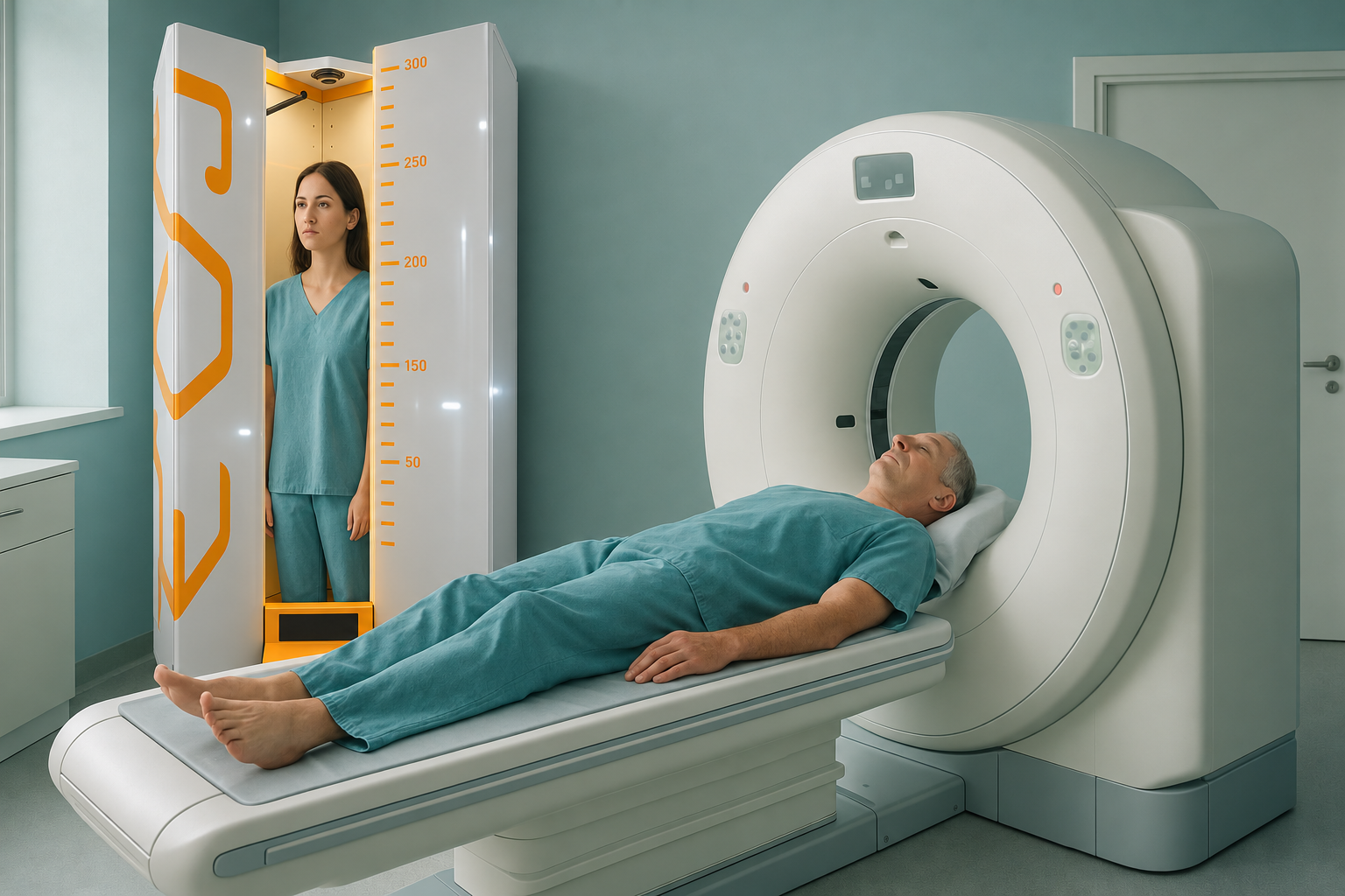
EOS Scan or CT Scan: How Do the Radiation Doses Compare?
An EOS scan uses about 90% less radiation than a standard X-ray, around ten times less. A CT scan gives a much higher dose again, because it takes many cross-sectional images. For questions about whole-body alignment, EOS gives detailed images of bones and joints at a small fraction of the radiation a CT scan would need.
Key points
- EOS uses about 90% less radiation than a standard X-ray, around ten times less.
- CT gives a much higher effective dose because it captures many cross-sectional slices.
- EOS scans you while you stand, so your bones and joints are shown carrying your real body weight.
- CT is still essential when your clinician needs detailed cross-sectional or soft-tissue bone images.
- Both follow the ALARA principle: use the lowest dose that answers the clinical question.
You may have been told you need a scan for your back, hip, knee or posture. If so, you may wonder how an EOS scan compares with a CT scan for radiation. It is a fair question. Knowing the difference in dose helps you and your clinician make an informed choice about the right scan for you.
How much radiation does an EOS scan use compared with a CT scan?
EOS uses about 90% less radiation than a standard X-ray, around ten times less. A CT scan of the same part of the body usually gives a much higher dose again. This is because it takes hundreds of cross-sectional images to build a detailed 3D picture. Published dose data varies with the body area and the scanner settings. But the overall pattern stays the same: EOS sits at the low end, a standard X-ray in the middle, and CT at the higher end.
The table below shows widely quoted effective dose ranges for common bone and joint scans. Exact figures depend on the equipment, the patient's size and the area scanned. So they are shown as rough ranges, not fixed numbers.
| Type of scan | Typical effective dose range | What it shows |
|---|---|---|
| EOS (full spine/lower limb) | Approximately 0.03 to 0.3 mSv | Full-length bone and joint alignment while standing, in 2D and 3D |
| Standard X-ray (same area) | Approximately 0.3 to 1.5 mSv | Bone detail of one area, usually lying down or standing |
| CT (spine or lower limb) | Approximately 2 to 20 mSv | Cross-sectional slices with fine bone detail and some soft-tissue detail |
These figures come from published research and manufacturer data. They are not exact for every patient. But they show why EOS is preferred when the clinical question is about alignment rather than the inner structure of the bone.
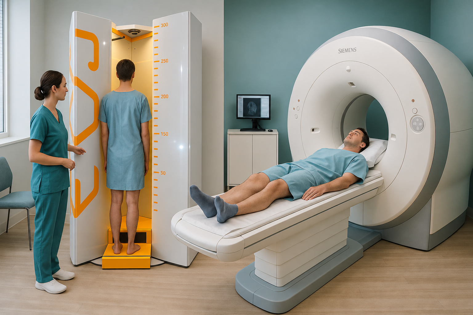
For a fuller look at how the three main types of scan compare, see our guide to EOS compared with X-ray, CT and MRI.
When is a CT scan the right choice instead of EOS?
CT is the right choice when your clinician needs detailed cross-sectional images of one area. It is excellent at showing fractures in complex bones, planning some surgeries, and assessing conditions such as femoral anteversion in adults through torsion measurements. Because CT captures thin slices, it can show bone detail that a single standing image cannot.
- You need the full-length alignment of your spine, hips and legs
- Your posture while carrying your body weight matters for the diagnosis
- You want the lowest practical radiation dose
- You may need follow-up scans over time
- A fracture needs precise cross-sectional detail
- Your surgeon needs rotational (torsion) measurements
- The inner structure of the bone is the main concern
- Your clinician has asked for a CT for a specific reason
Both scans follow the ALARA principle (As Low As Reasonably Achievable). The aim is always to use the scan that answers the clinical question with the least radiation needed. If your question is about overall alignment, EOS will usually answer it with a far lower dose than CT.
Why does a standing scan change the picture?
A standing scan changes the picture because gravity and body weight affect how your bones, joints and spine line up. When you lie down for a CT scan, your muscles relax, your curves flatten, and small alignment problems can disappear. The body is a connected chain from your feet to your head. A tilt in one area often makes another area compensate. This is why pain often shows up away from its actual cause.
An EOS scan captures the whole skeleton while you stand naturally. So the image shows how your body really works when you are upright, walking and carrying load. For questions about alignment, this standing view often shows what a lying-down CT, X-ray or MRI misses.
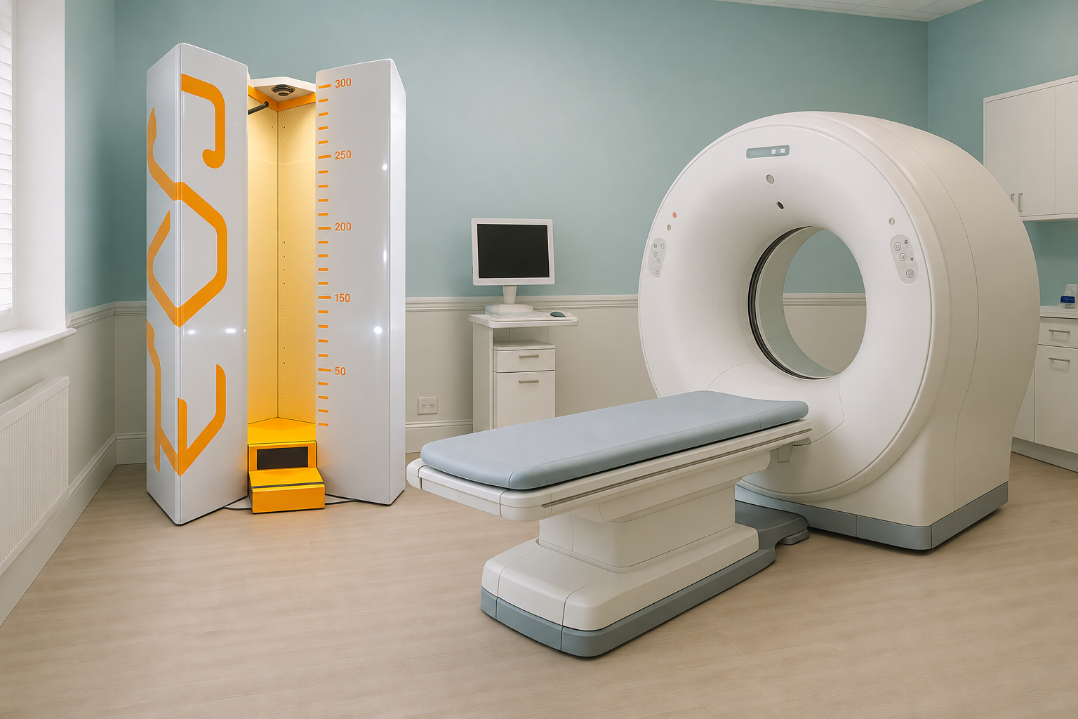
Read more about EOS scan safety and radiation to see how the low dose is achieved.
How do I book a low dose standing scan at ScanAlign?
You do not need a referral from your doctor. You can book directly with us, even from abroad, and the process is simple.
Free video consultation
Speak with our specialist radiographer from home. Talk through your symptoms and confirm that an EOS scan is the right next step for you.
Come for your scan
Visit ScanAlign, low dose standing imaging at 19 Harley Street, London. The scan takes just a few minutes while you stand naturally.
Receive your report
A specialist radiology report sets out your alignment findings. You can share it with your doctor, surgeon or physiotherapist back home.
Next steps
Use the report to guide your talks with your treating clinician about what to do next.
Want to find out if a standing EOS scan suits your situation?
Book a free video consultationA note about MRI. EOS shows bones, joints and alignment very clearly. For soft tissue, discs or nerves, MRI is usually the better tool. If your clinician suspects a disc problem or a pressed nerve, MRI may be advised alongside EOS or instead of it. The two scans answer different questions and often work well together.
Before you start treatment, or change the one you are on, it is worth getting a detailed diagnosis. Knowing whether the cause sits in your pelvis, your legs or your spine changes what will actually help.
Frequently asked questions
EOS uses about 90% less radiation than a standard X-ray, around ten times less. CT uses much more. For questions about alignment, EOS gives the information needed at a much lower dose. Both are considered safe when there is a clear clinical reason for them.
Not always. CT gives detailed cross-sectional images that EOS does not. If your clinician needs to see inside a bone or assess a complex fracture, a CT scan may still be needed. EOS is best for whole-body alignment seen while standing.
ALARA stands for As Low As Reasonably Achievable. It is the guiding principle in radiology: always use the lowest radiation dose that still answers the clinical question. EOS fits well with ALARA because it gives very low doses for full-body skeletal imaging.
The scan itself takes only a few minutes. You stand still inside the scanner while it takes the images. The whole appointment, including preparation, is a short visit.
No. ScanAlign accepts patients who refer themselves. You can book directly through a free video consultation to discuss whether the scan is right for you.
A CT torsion study measures the rotation of bones such as the femur or tibia. CT is usually used for this because it captures cross-sectional slices. EOS can give some rotational information through its 3D reconstruction. But detailed torsion measurements may still need CT, depending on the clinical question.
ScanAlign is a private self-pay clinic. The price for patients travelling from abroad is confirmed during the free video consultation, after which you can book a low dose standing scan.
A single CT scan is generally considered safe. The risk from any one scan is very small. However, repeated scans add up over a lifetime. This is why clinicians use CT only when the benefit for diagnosis outweighs the radiation exposure. For alignment questions where CT-level detail is not needed, a lower-dose option such as EOS is often preferable.
As with any X-ray-based scan, EOS is generally avoided during pregnancy unless it is clinically essential. If you are or might be pregnant, tell the team before your appointment so they can advise you.
When you stand, gravity loads your spine, hips and legs the way they work in daily life. Lying down relaxes the muscles and can hide tilts, shifts and compensations that only appear under load. A standing scan shows your real alignment, which often explains symptoms that lying-down scans cannot.
Wondering if EOS is the right scan for you?
Speak with our specialist radiographer in a free video consultation, from wherever you are. No referral needed, no obligation.
Book a free video consultationWritten by Abbas Dhami (Specialist Diagnostic Radiographer)
Sources
- Deschênes S, Charron G, Beaudoin G, et al. "Diagnostic imaging of spinal deformities: reducing patients' radiation dose with a new slot-scanning X-ray imager." Spine, 2010. (EOS dose data)
- Public Health England. "Patient dose information: guidance." GOV.UK, 2011. (UK national reference doses for X-ray and CT)
- American College of Radiology. "ACR Appropriateness Criteria: Radiation Dose Assessment." ACR.org. (ALARA principle and justifying a scan)
This page is general information and not a diagnosis.
ScanAlign is the trading name of CoreMed Solutions Ltd and operates under The Harley Street Hospital's CQC licence.