
How Is an EOS Scan Different From a Standard X-ray?
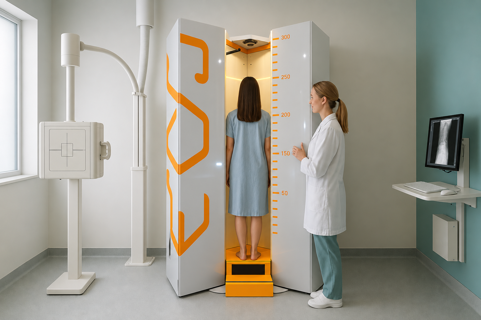
An EOS scan takes front and side images of your whole body at the same time, while you stand. It uses about 90% less radiation than a standard X-ray, around ten times less. Because your body is bearing its own weight, the images show your true alignment under gravity. A normal X-ray taken lying down cannot reliably do this.
Key points
- EOS uses slot-scanning technology to image your whole skeleton in one pass, with no stitching or magnification errors.
- It gives about 90% less radiation than a standard X-ray, around ten times less.
- You stand naturally during the scan, so the images show your real, weight-bearing alignment.
- Calibrated measurements help specialists plan treatment with greater precision.
- EOS shows bones and joints. For soft tissue, discs or nerves, MRI is usually the better tool.
What is an EOS scan and how does it work?
An EOS scan is a type of biplanar X-ray. It takes a front image and a side image at the same time. A standard X-ray uses a single flash of radiation. EOS instead uses two narrow slot-scanning beams that move from head to toe in about 15 seconds. This captures your full skeleton in one continuous pass.
The result is a life-size, calibrated image. There is no magnification, and separate images do not need to be stitched together. Your specialist can take precise measurements straight from the scan. If needed, the data can also be used to build a 3D model of your skeleton.
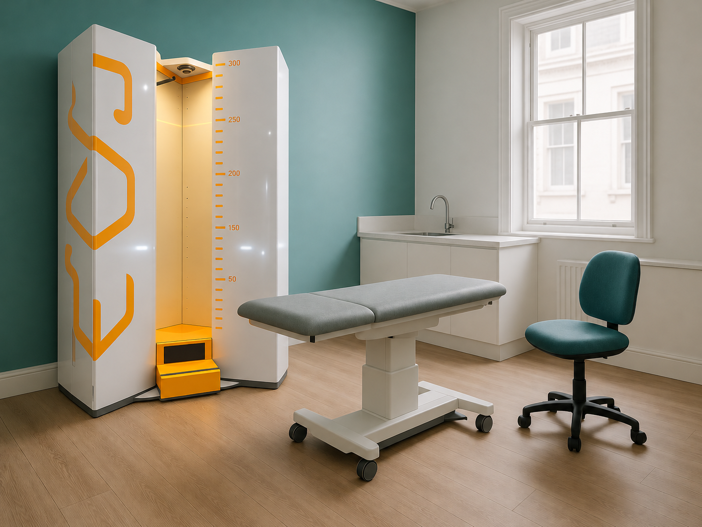
At ScanAlign, standing EOS imaging in London, this technology is used to check whole-body alignment from your feet to your skull. It gives specialists a complete picture of how your skeleton balances under load.
How does EOS compare to a standard X-ray?
EOS differs from a normal X-ray in several important ways. The table below sums up the main differences.
| Feature | Standard X-ray | EOS Scan |
|---|---|---|
| Position | Often lying down or sitting | Standing, weight-bearing |
| Coverage | One area at a time | Full body in a single pass |
| Views taken | One view per exposure | Front and side at the same time |
| Radiation dose | Standard dose | About 90% less (around ten times less) |
| Image stitching | Several images stitched together for full-length views | No stitching needed, one continuous image |
| Magnification | Some magnification distortion | Calibrated, life-size, no magnification |
| 3D model | Not available | Available from the biplanar data |
A standard X-ray is excellent for looking at one area, such as a broken wrist or a chest infection. But when the question is about alignment, posture or how one part of your body relates to another, it can miss the full picture. This is because it covers a small area and is often taken lying down. That is where an EOS standing X-ray adds value.
Why does standing up during imaging matter?
Standing up changes everything. When you lie down for a standard X-ray, your muscles relax and gravity no longer presses on your joints. A curve in your spine may flatten out. A tilt in your pelvis may disappear. The image looks normal, even though you feel pain every time you stand or walk.
A standing weight-bearing X-ray captures your skeleton as it really works. Your body is a connected chain from your feet to your head. If one area is out of alignment, other areas make up for it. Pain often shows up away from its true cause, in your knee rather than your hip, or in your lower back rather than your pelvis.
Because EOS images your full body in one standing pass, it can show these compensation patterns. For example, a full spine X-ray taken with EOS shows how your neck, mid-back, lower back and pelvis relate to each other in a single image, all under real weight-bearing conditions.
Wondering whether your pain might be caused by an alignment problem that standard imaging has missed?
Book a free video consultationWhat happens when you book an EOS scan?
The process is simple. You do not need a referral from your doctor to book an EOS scan.
Free video consultation
You speak with a specialist radiographer. They listen to your symptoms and confirm whether an EOS scan is the right next step for you.
Come for your scan
You come to 19 Harley Street, London. The scan itself takes about 15 seconds. You stand still in the EOS booth while the system takes your images.
Expert report
A specialist radiologist studies your images and writes a detailed report on your alignment from head to toe.
Results review
You receive your report and images. You can share these with your consultant, surgeon, physiotherapist or any other clinician treating you.
When is MRI a better choice than EOS?
MRI is usually the better tool when the question is about soft tissues rather than alignment. EOS is excellent at showing bones, joints and the way your skeleton lines up under load. It does not image soft tissue in detail.
EOS and MRI do different jobs. EOS shows bones, joints and whole-body alignment under real weight-bearing conditions. For soft tissue problems, disc herniations or nerve compression, MRI is usually the better tool. Many patients benefit from both, depending on the clinical question.
Before you start treatment, or change the one you are on, it is worth getting a detailed diagnosis. Knowing whether the cause sits in your pelvis, your legs or your spine changes what will actually help.
Frequently asked questions about EOS vs X-ray
EOS uses about 90% less radiation than a standard X-ray, around ten times less. This makes it a lower-dose option. That is especially useful when full-body or repeat imaging is needed.
No. You can book directly yourself. You start with a free video consultation to check that EOS imaging is right for you.
The scan itself takes about 15 seconds. Your whole appointment will be a little longer, to allow time for positioning and any questions you have.
EOS captures your whole skeleton in one standing pass, with front and side views at the same time. This shows whole-body alignment and compensation patterns that X-rays of one area, taken lying down, can miss.
No. They answer different questions. EOS is for bones, joints and alignment. MRI is for soft tissue, discs and nerves. Your clinician may recommend one or both, depending on your symptoms.
No. A CT scan uses several rotating X-ray beams and gives a much higher radiation dose. EOS uses two low-dose slot-scanning beams and images you standing up rather than lying in a tube.
Biplanar means two planes. EOS takes a front-to-back image and a side image at the same time. This gives two views in a single exposure, instead of needing separate X-rays.
ScanAlign is a private, self-pay clinic. The price for patients travelling from abroad is confirmed during your free video consultation, along with exactly what is included.
When you lie down, gravity stops pressing on your joints and your muscles relax. Curves, tilts and imbalances that cause symptoms when you stand can flatten out and look normal on an image taken lying down.
ScanAlign offers EOS standing imaging at 19 Harley Street, London. EOS is uncommon in the UK, but it is not only available at ScanAlign. Other clinics, including some on Harley Street, also offer EOS imaging.
See the difference for yourself
Find out whether a standing EOS scan could show what other imaging has missed. Start with a free, no-pressure video consultation.
Book a free video consultationWritten by Abbas Dhami (Specialist Diagnostic Radiographer)
Sources
- Deschênes S, Charron G, Beaudoin G, et al. Diagnostic imaging of spinal deformities: reducing patients' radiation dose with a new slot-scanning X-ray imager. Spine. 2010;35(9):989-994.
- Illés T, Somoskeöy S. The EOS imaging system and its uses in daily orthopaedic practice. International Orthopaedics. 2012;36(7):1405-1411.
- Royal College of Radiologists. iRefer: Making the best use of clinical radiology. rcr.ac.uk.
This page is general information and not a diagnosis.
ScanAlign is the trading name of CoreMed Solutions Ltd and operates under The Harley Street Hospital's CQC licence.