
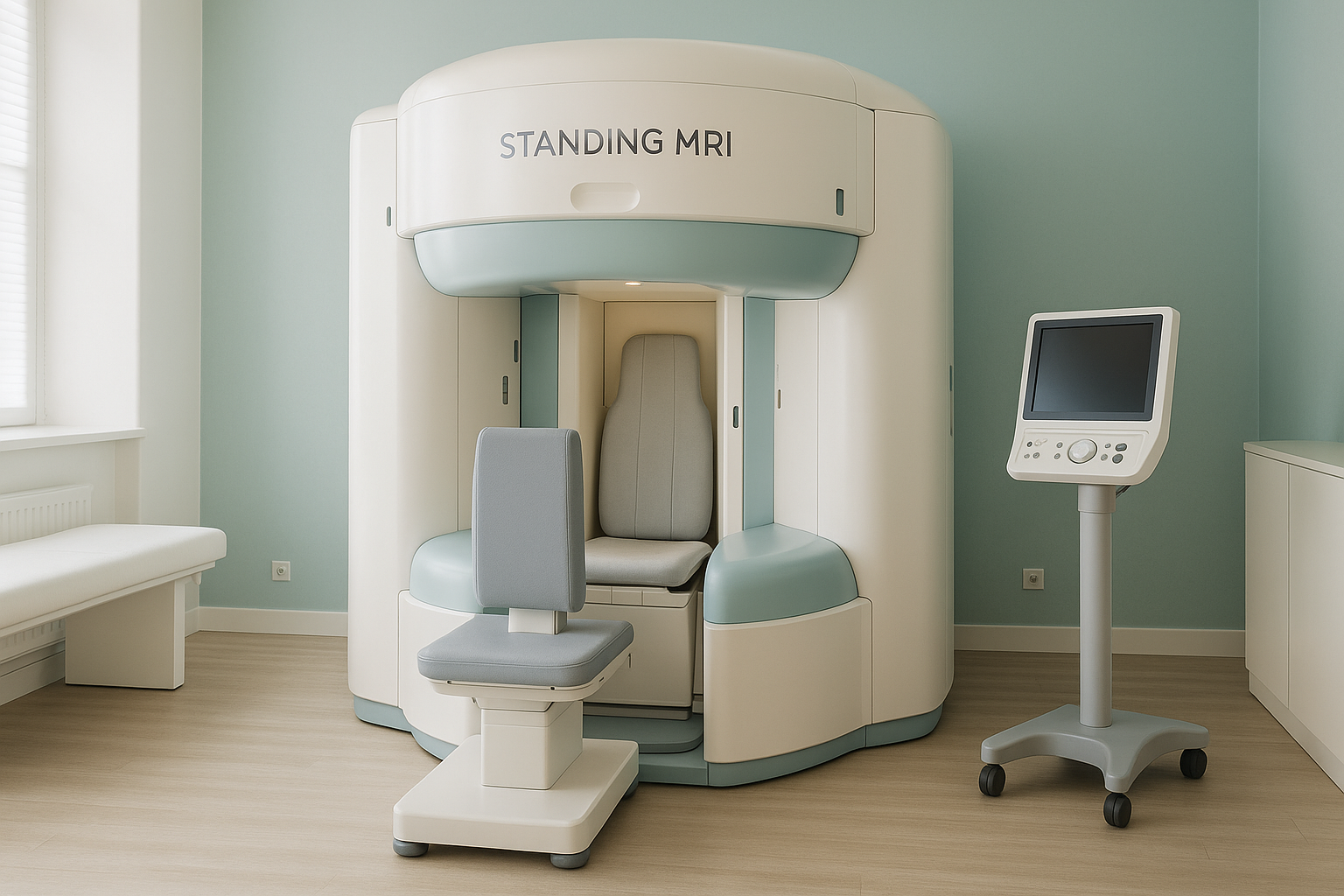
EOS or Standing MRI: Which Upright Scan Do You Actually Need?
A standing MRI uses magnets to show soft tissue (discs, nerves, ligaments) while you are upright. An EOS scan uses very low-dose X-rays to capture your full skeletal alignment from head to feet in one image. The two scans answer different questions. So the right choice depends on what your clinician needs to see.
Key points
- Standing MRI is excellent for soft tissue under load, such as disc bulges that only appear when you are upright.
- EOS is excellent for whole-body skeletal alignment. It shows how your spine, hips, knees and ankles relate to each other under gravity.
- The two scans complement each other. Neither replaces the other.
- Standing MRI scans are slower than conventional MRI, and less widely available in the UK.
- An EOS scan delivers about 90% less radiation than a standard X-ray, around ten times less.
What does a standing MRI show compared to EOS?
A standing MRI (also called an upright MRI or positional MRI) shows soft tissue, including discs, nerves, muscles and ligaments, while your body carries its own weight. This matters because some disc bulges or nerve compressions only appear when gravity is acting on the spine. A conventional MRI, done lying down, can miss these positional findings. Read more about weight-bearing versus lying imaging.
An EOS standing X-ray, on the other hand, captures the whole skeleton from head to feet in a single standing image. It shows bone position, joint angles and overall sagittal balance. It does not show soft tissue in any detail. But it gives a complete map of how your skeleton is aligned when it is carrying real weight.
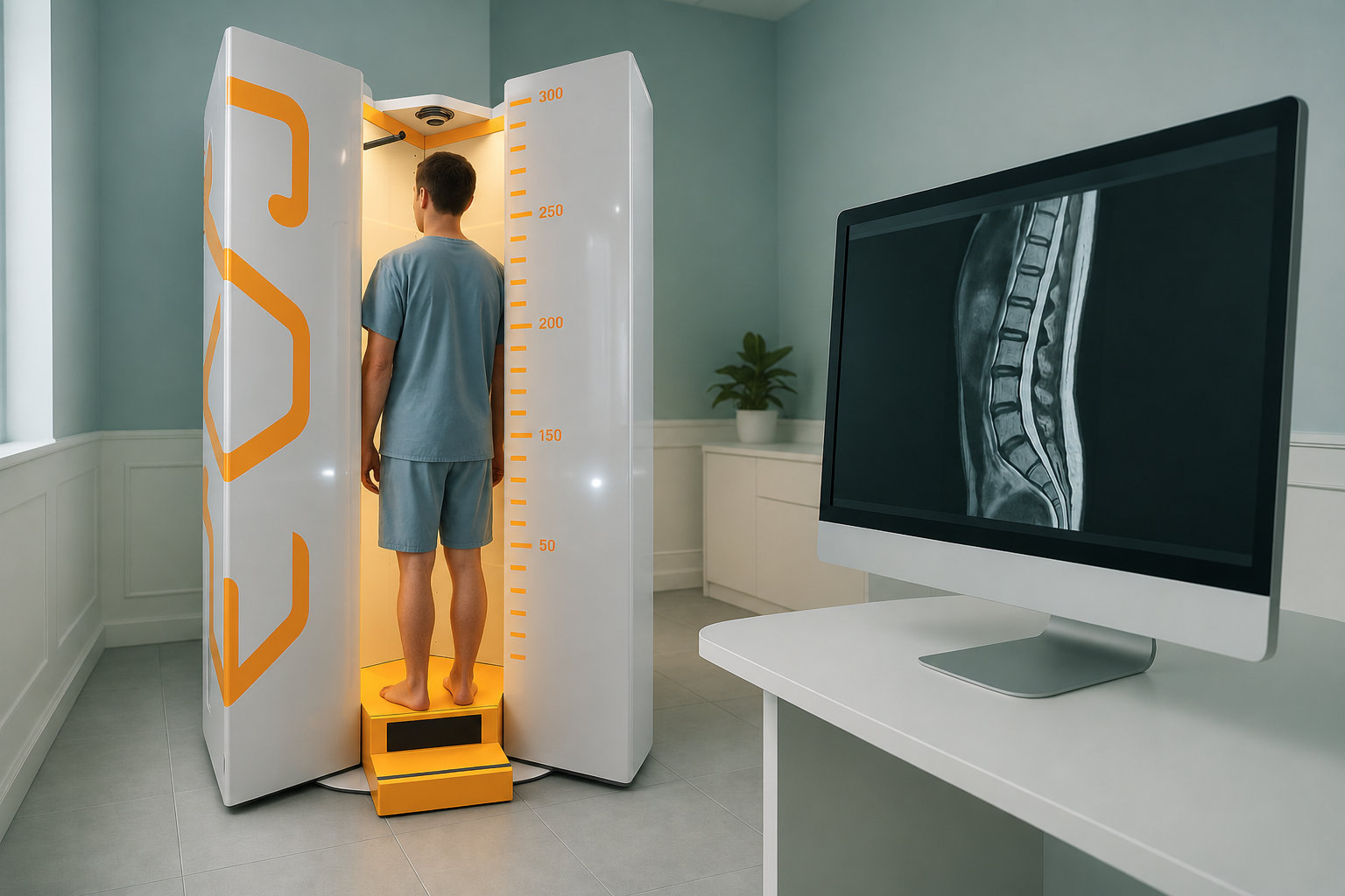
How do EOS and upright MRI compare side by side?
The table below sums up the practical differences. Both scan you standing up. But they use very different technology and answer different clinical questions.
| Feature | EOS standing scan | Standing / upright MRI |
|---|---|---|
| What it images | Bones, joints, skeletal alignment | Soft tissue: discs, nerves, ligaments, muscles |
| Coverage | Whole body, head to feet, in one exposure | One focused area (e.g. lower back, one knee) |
| Radiation | About 90% less than a standard X-ray | None (uses magnets, no radiation) |
| Scan time | Under a minute for the exposure | Typically 30 to 60 minutes per area |
| Magnet strength | Not applicable | Usually 0.5 T or lower (conventional MRI is 1.5 to 3 T) |
| Image quality trade-off | Excellent bone detail at very low dose | Lower field strength can mean less fine soft-tissue contrast than conventional MRI |
| Availability in the UK | Uncommon, but available at select private clinics | Very limited, only a handful of sites across the UK |
| Best for | Alignment assessment, surgical planning, scoliosis, leg length, joint angles | Positional disc bulges, nerve compression under load, some soft-tissue injuries |
Which standing scan is right for your problem?
The answer depends on the question your clinician is trying to answer. Here is a simple way to think about it.
- You need the full picture of how your spine, pelvis, hips, knees and ankles line up
- A surgeon is planning a spinal fusion, hip replacement or knee realignment
- You have unexplained pain that moves around, suggesting one area is compensating for another
- You want the lowest possible radiation dose for an overview of your skeleton
- You need to see a disc bulge or nerve compression that only appears when you are upright
- A conventional MRI done lying down showed nothing, yet symptoms continue when you stand or sit
- Your clinician specifically needs soft-tissue detail while your body is carrying weight
- You cannot have any radiation exposure at all
In many complex cases, both scans add value. An EOS image shows the mechanical "big picture". A standing MRI fills in the soft-tissue detail at the problem level. Your clinician can then put both sets of information together for more complete planning.
A note on MRI. EOS is designed for bones, joints and alignment. For soft tissue, discs or nerves, MRI (conventional or upright) is usually the better tool. If your question is purely about soft tissue, MRI is likely the place to start. If it is about alignment and posture, EOS is built for that job.
Not sure which scan you need? We can help you work that out before you commit to anything, including travel to London.
Book a free video consultationWhat are the practical trade-offs of an upright MRI?
Standing MRI scanners usually use a lower magnetic field strength, often around 0.5 Tesla. A conventional tunnel MRI uses 1.5 or 3 Tesla. That lower field can mean less contrast between soft tissues, so very small lesions are harder to spot. Scan times are also longer, which can be tiring if you are already in pain.
Weight-bearing MRI is also far less widely available than conventional MRI. Only a handful of clinics across the UK offer it at present.
EOS, by contrast, takes under a minute for the exposure. It gives a full-length standing image that is ready for analysis quickly. The trade-off is that it shows bones and alignment, not soft tissue. That is why the two technologies complement each other rather than compete. See what ScanAlign standing EOS imaging involves in practice.
Booking an EOS scan at ScanAlign
You do not need a referral from your doctor. If you want to understand your alignment, you can book a standing scan directly. If your clinician thinks you also need a standing MRI, we will tell you honestly during your free consultation. The price for patients travelling from abroad is confirmed during the free video consultation.
Before you start treatment, or change the treatment you are on, it is worth getting a detailed diagnosis. Knowing whether the cause sits in the pelvis, the legs or the spine changes what will actually help.
Frequently asked questions
Yes. "Standing MRI", "upright MRI" and "positional MRI" all describe the same idea: an MRI scanner that images you while you are upright rather than lying in a tunnel. The exact design varies between manufacturers.
No. MRI uses strong magnets and radio waves, so there is no ionising radiation at all. EOS does use X-rays, but at about 90% less radiation than a standard X-ray, around ten times less.
No. EOS shows bones, joints and skeletal alignment. MRI shows soft tissue such as discs, nerves and ligaments. They answer different clinical questions and often complement each other.
Standing MRI is available at a small number of private clinics in London. It is harder to find than conventional MRI. ScanAlign offers EOS standing imaging, not standing MRI. During a free consultation, we can help you decide which type of scan suits your needs.
We do not offer standing MRI at ScanAlign, so we cannot quote MRI prices. For an EOS scan, the price for patients travelling from abroad is confirmed during the free video consultation.
When you lie down, gravity no longer compresses your spine. Discs can pull back slightly and a bulge may shrink for a while. Standing loads the spine with your full body weight. This can push the disc material outward and press on a nerve. A weight-bearing scan captures how your spine behaves in real life.
No. ScanAlign accepts self-referrals. You can book a free video consultation directly, with no referral letter needed.
Most standing MRI scanners use a lower field strength (often 0.5 T) than conventional scanners (1.5 to 3 T). This can reduce fine soft-tissue contrast. For many positional questions, the trade-off is worthwhile. But for very detailed soft-tissue assessment, conventional MRI may still be preferred.
The X-ray exposure itself takes under a minute. Including positioning and preparation, you will be in the scanning room for only a short time. A standing MRI, by comparison, can take 30 to 60 minutes per body area.
Yes, and in complex cases this combination can be very informative. The EOS gives the alignment overview. The standing MRI adds soft-tissue detail at the problem level. Your clinician can advise whether both are needed.
Not sure which scan you need?
Talk it through with a specialist radiographer by video, before you plan any travel. No charge, no commitment.
Book a free video consultationAuthor: Abbas Dhami (Specialist Diagnostic Radiographer)
Sources
- Jinnas K, Sward A, Hansson T, et al. "Positional MRI of the lumbar spine." European Spine Journal, 2006;15(Suppl 3):S361-S368.
- Deschênes S, Charron G, Beaudoin G, et al. "Diagnostic imaging of spinal deformities: reducing patients' radiation dose with a new slot-scanning X-ray imager." Spine, 2010;35(9):989-994.
- The Royal College of Radiologists. iRefer Guidelines: Making the Best Use of Clinical Radiology. RCR, London (current edition).
This page is general information and not a diagnosis.
ScanAlign is the trading name of CoreMed Solutions Ltd and operates under The Harley Street Hospital's CQC licence.