
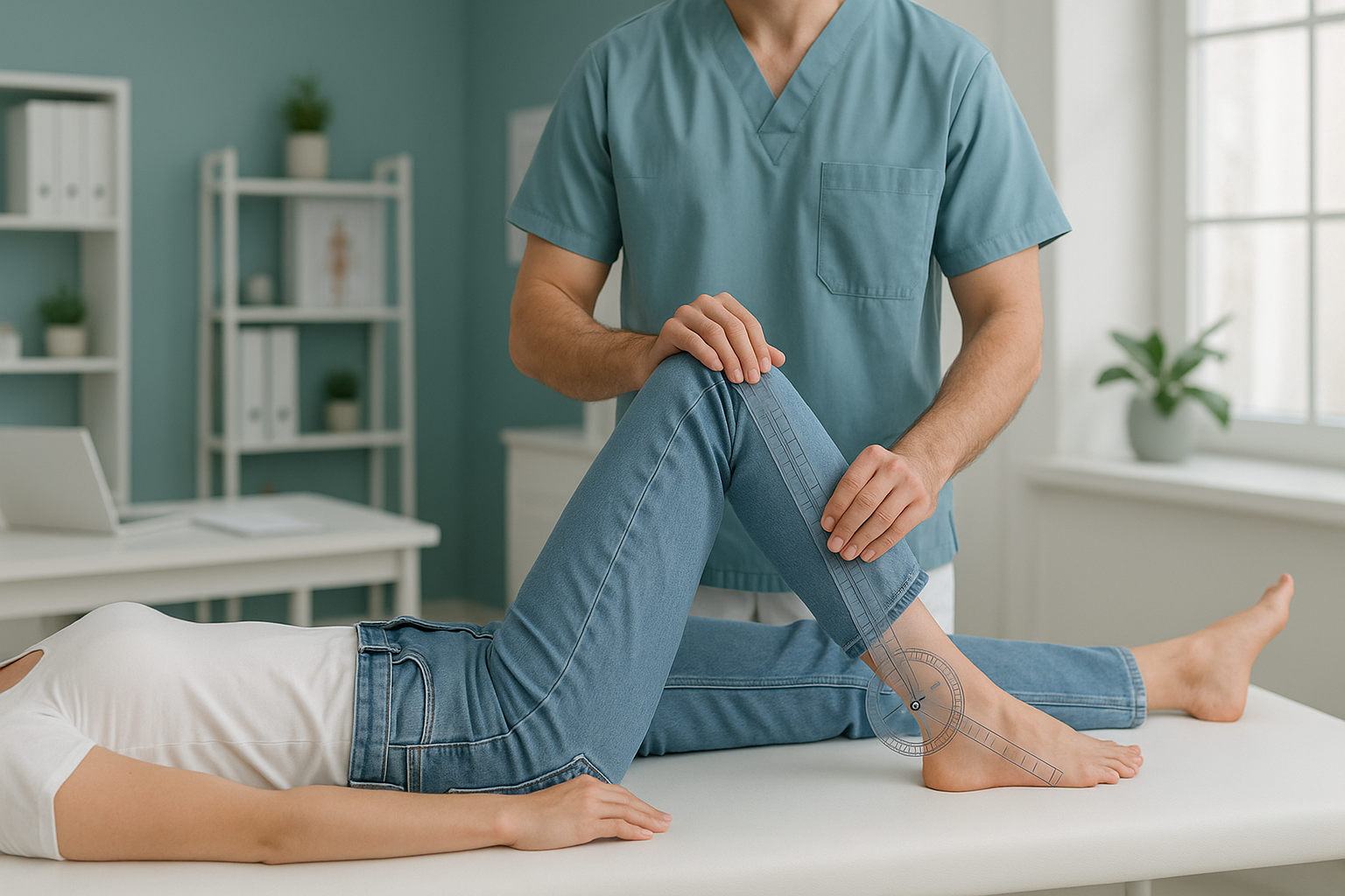
How Is Femoral Torsion Measured Without a CT Scan?
Femoral anteversion and torsion can be measured accurately with a low dose biplanar EOS scan instead of a CT scan. The EOS system takes two standing images at the same moment. It then builds a 3D model of your femur. This gives your surgeon the version angle they need, with about 90% less radiation than a standard X-ray, around ten times less.
Key takeaways
- Femoral torsion describes how much your thigh bone is twisted along its length.
- CT has been the usual way to measure it, but it gives a fairly high radiation dose.
- EOS biplanar imaging makes a 3D model of the whole leg while you stand, using about 90% less radiation than a standard X-ray.
- A standing measurement shows how torsion affects your alignment when your legs are carrying your real body weight.
- The results can guide decisions about physiotherapy, gait retraining, or surgery planning such as derotation osteotomy.
What is femoral anteversion and why does it matter?
Femoral anteversion is the natural forward twist of your thigh bone (femur) between the hip joint and the knee. Everyone has some of this twist. When it falls outside the usual range, clinicians call it excessive anteversion. If the bone twists the other way, they call it retroversion. You can read more about the difference between anteversion versus retroversion.
Too much or too little twist changes the way your leg sits in the hip socket. It can cause in-toeing (feet pointing inward when you walk), hip pain during activity, or limited inward and outward rotation of the hip. Over time, it may add to cartilage wear, or to problems at the knee or lower back as the body compensates. Understanding femoral anteversion in adults is an important step in working out why your symptoms keep going.
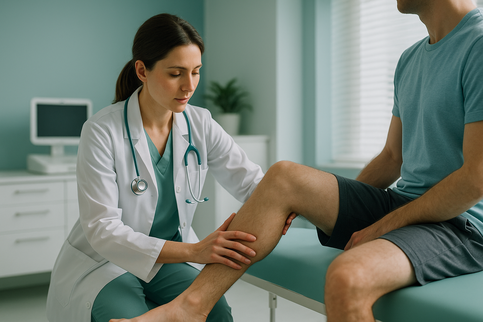
Why is CT the usual way to measure femoral torsion?
CT has been the standard test for measuring femoral version. It takes cross-sectional slices through the hip and knee, so the radiologist can work out the twist angle precisely. It works well for the measurement itself, but it has two important drawbacks.
First, a CT rotational profile of both legs involves a meaningful dose of ionising radiation aimed at the pelvis and knees. Many people need this measurement repeated over time, and younger adults may need it before surgery. So the dose adds up.
Second, CT is done lying down. Your muscles relax, gravity no longer loads your joints, and the image does not show how your bones and joints really line up when you stand and walk. That is a big gap, because the whole point of the measurement is to understand how torsion affects your movement in daily life.
How does EOS measure femoral torsion without CT?
EOS is a biplanar imaging system. It takes a front view and a side view at the same time, in one standing scan that lasts about 20 seconds. Specialist software then uses those two views to build a calibrated 3D model of your skeleton.
From that 3D model, the version angle of each femur can be worked out. So can tibial torsion, knee alignment, and the overall rotation of the leg from hip to ankle. Because you stand in your natural posture, the measurements show your real weight-bearing alignment, not a relaxed position on a scanner table.
The radiation dose is about 90% less than a standard X-ray, around ten times less. For a scan that covers both legs from the pelvis to the feet, that is a large difference compared with CT. You can read a detailed breakdown of EOS compared with X-ray, CT and MRI.
How does EOS compare with CT for measuring torsion?
EOS and CT both give accurate version angles. They differ in radiation dose, in how the patient is positioned, and in how much information they give in a single scan.
| Factor | CT rotational profile | EOS biplanar scan |
|---|---|---|
| Patient position | Lying down | Standing, weight-bearing |
| Radiation dose | Higher | About 90% less than a standard X-ray |
| 3D model | Yes (from many slices) | Yes (from two views taken at the same time) |
| Full-leg alignment | Not in one scan | Pelvis to feet in one image |
| Tibial torsion | Needs extra slices | Included in the same scan |
| Soft tissue detail | Good | Not applicable (bones and joints only) |
When MRI is the better choice: EOS shows bones, joints and alignment. If your clinician needs to see soft tissue such as cartilage, the labrum, muscles or nerves, MRI is usually the better test. The two scans answer different questions and often work well together.
What are the steps to get a femoral torsion measurement at ScanAlign?
You do not need a referral from your doctor. The process is simple, and you can contact us yourself at any stage.
Free video consultation
A short call with a specialist radiographer to talk about your symptoms and confirm whether an EOS scan is the right next step for you.
Come for your scan
You stand inside the EOS unit at our clinic on Harley Street in London. The scan takes about 20 seconds and covers both legs from the pelvis to the feet.
3D model and analysis
The biplanar images are used to build a 3D model of your legs. Femoral version, tibial torsion and your full rotational profile are all worked out from this model.
Your report
You receive a detailed report on your rotational profile and alignment findings. This is exactly what a surgeon or physiotherapist needs for planning. Find out more about what an EOS scan report shows.
Who should consider an EOS torsion measurement?
A standing rotational profile is useful for anyone with unexplained hip, knee or walking problems where torsion may play a part. It is not the right test for every situation.
- In-toeing or out-toeing that continues into adult life
- Hip pain with limited inward or outward rotation
- Planning before derotation osteotomy surgery
- Follow-up measurement with lower radiation than CT
- Femoral and tibial torsion both need to be assessed
- Soft tissue injury (labral tear, muscle strain). MRI is better.
- A new fracture or injury. You need the emergency department or urgent imaging first.
- A concern about infection or a tumour. CT or MRI with contrast is more suitable.
Before you start treatment, or change the one you are on, it is worth getting a detailed diagnosis. Knowing whether the cause sits in the pelvis, the legs or the spine changes what will actually help you.
Not sure whether an EOS scan is right for you? Start with a free video consultation. A specialist radiographer will talk through your symptoms and give you honest advice.
Book a free video consultationFrequently asked questions
Femoral anteversion is the forward twist of your thigh bone between the hip and the knee. A certain amount is normal. When the angle is unusually large or small, it can affect hip rotation, the way you walk, and how comfortable your joints feel.
Traditionally it is measured with a CT scan that takes cross-sectional images of the hip and knee. EOS biplanar imaging is another option. It builds a 3D model from two standing views, so the version angle can be worked out with much less radiation.
Published research shows strong agreement between EOS 3D models and CT-based measurements of femoral version. Your clinician can discuss whether EOS suits your specific clinical question.
An EOS scan uses about 90% less radiation than a standard X-ray, around ten times less. This matters most for full-leg scans that cover the pelvis, thighs and knees.
Yes. EOS captures the full leg from the pelvis to the feet in one standing scan. So femoral version and tibial torsion are both measured from the same 3D model.
No. You can contact us directly. We recommend starting with a free video consultation, so a specialist radiographer can confirm that EOS is the right scan for your needs.
The price for patients travelling from abroad is confirmed during the free video consultation, and you can book a torsion measurement scan online.
When you lie down, your muscles relax and gravity no longer loads your joints. Standing imaging shows how torsion, alignment and rotation work together under the same forces your body carries every day. This gives a more clinically useful picture.
Yes. The 3D rotational profile from an EOS scan gives the precise version angles a surgeon needs to plan a derotation osteotomy. Many surgeons now accept EOS data instead of CT for this purpose.
ScanAlign is based at 19 Harley Street, London. We are a private, self-pay clinic offering ScanAlign, low dose standing imaging to assess whole-body alignment.
Get your torsion measured with less radiation
A free video consultation is the first step. We will tell you honestly whether an EOS scan is the right choice for you.
Book a free video consultationWritten by Abbas Dhami (Specialist Diagnostic Radiographer)
Sources
- Guenoun B, et al. Reliability of a new method for lower-extremity measurements based on stereoradiographic three-dimensional reconstruction. Orthopaedics & Traumatology: Surgery & Research, 2012;98(5):506-513.
- Buck FM, et al. Femoral and tibial torsion measurements with 3D models based on low-dose biplanar radiographs in comparison with standard CT measurements. American Journal of Roentgenology, 2012;199(5):W607-W612.
- EOS imaging clinical evidence library. EOS imaging (now part of Alphatec Holdings). Available at: eos-imaging.com.
This page is general information and not a diagnosis.
ScanAlign is the trading name of CoreMed Solutions Ltd and operates under The Harley Street Hospital's CQC licence.