
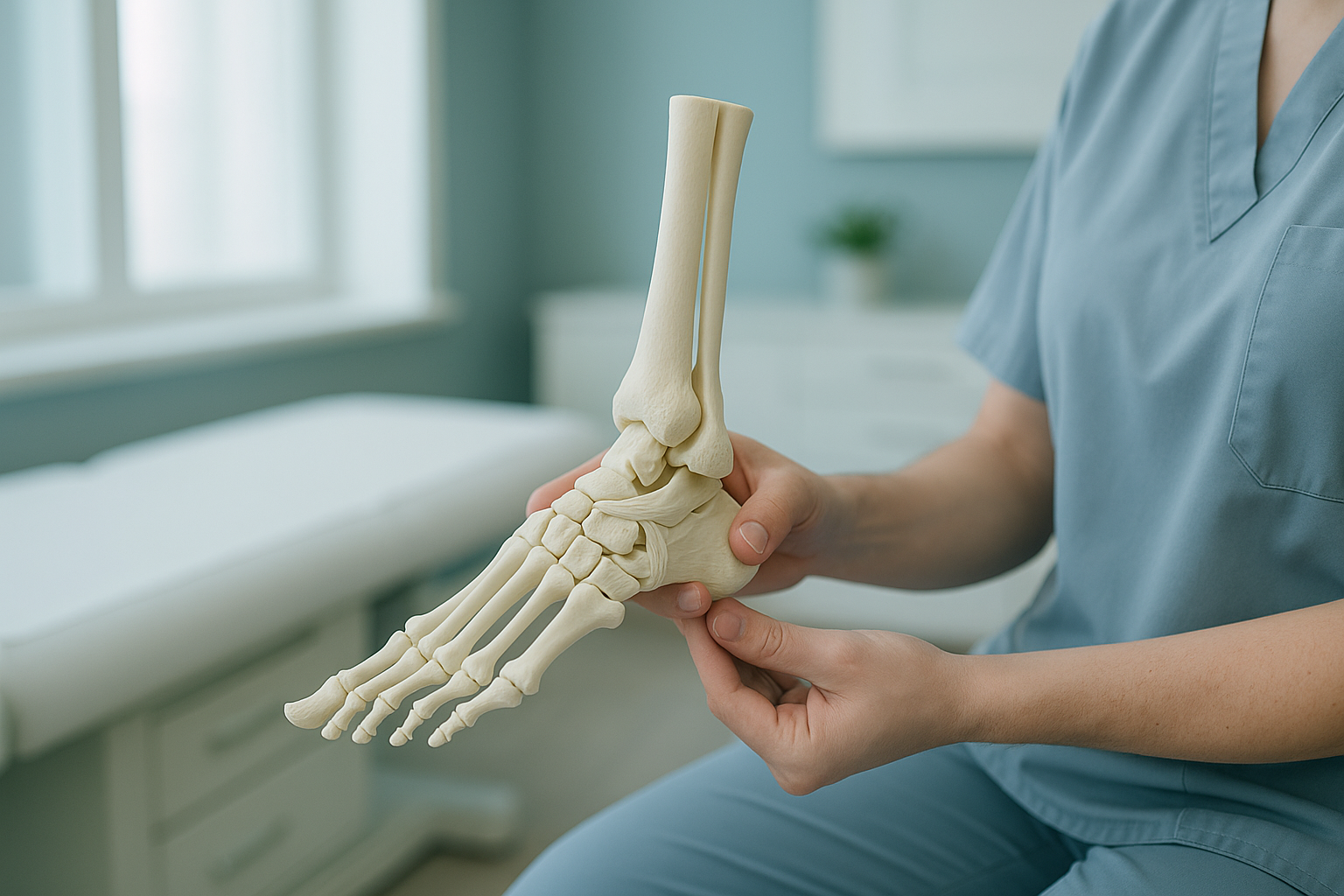
Foot and Ankle Anatomy: What Every Bone and Ligament Actually Does
Your foot has 26 bones, over 30 joints and more than 100 ligaments and tendons that work together. The ankle ligaments keep your ankle stable. The arches absorb shock. The bones pass your whole body weight down to the ground. When any part is out of alignment, pain can show up in the foot, knee, hip or back.
Key takeaways
- Each foot has 26 bones, split into three regions: hindfoot, midfoot and forefoot.
- Ankle ligaments on the outer and inner side stop the joint from rolling.
- The medial arch is your main shock absorber. The plantar fascia supports it.
- Misalignment in the foot can cause compensatory pain higher up the body.
- Standing, weight-bearing imaging shows how these parts behave under real load.
How many bones are in the foot and what do they do?
Each foot has 26 bones. So both feet together hold nearly a quarter of all the bones in your body. They are grouped into three regions that work as a team.

Hindfoot
The calcaneus (heel bone) is the largest bone in the foot. It absorbs the first impact each time your foot hits the ground. The talus sits on top of it and forms the ankle joint with your lower leg bones. Together they make the subtalar joint, which lets your foot tilt inward and outward.
Midfoot
Five bones, including the navicular, cuboid and three cuneiforms, bridge the gap between the heel and the forefoot. They lock together into a rigid lever when you push off while walking. The midfoot is where the arch gets its shape.
Forefoot
Five metatarsals fan out towards your toes. Fourteen smaller phalanges make up the toes themselves. The metatarsal heads carry a large share of your weight during the push-off phase of each step.
What are the main ankle ligaments and why do they matter?
Ankle ligaments are tough bands of tissue that join bone to bone. They hold the joint together but still allow movement. The ankle has ligaments on both sides, and each group resists a different direction of roll or twist.
Three ligaments, including the ATFL (anterior talofibular ligament), stop the ankle from rolling outward. The ATFL is the most commonly sprained ligament in the body.
A thick, fan-shaped ligament on the inside of the ankle. It resists the foot rolling inward and is stronger than the outer ligaments, so inner ankle sprains are less common.
These join the two lower leg bones (tibia and fibula) just above the ankle joint. A "high ankle sprain" injures this group and usually takes longer to heal.
When ankle ligaments are stretched or torn, the joint can become unstable. Over time, repeated instability changes how weight spreads across the foot. This can lead to problems such as peroneal tendonitis on the outer side of the ankle.
What does the arch of the foot actually do?
The arch of the foot works like a spring. It absorbs shock when you land and stores energy to help push you forward. You actually have three arches, but the medial (inner) arch matters most for carrying weight.

The plantar fascia, a thick band of connective tissue along the sole, supports the medial arch like a bowstring. If the arch flattens too much (overpronation), the plantar fascia is overstretched, and your ankle, knee and hip have to compensate. If you have ongoing foot pain linked to flat feet or rolling ankles, reading about orthotics and overpronation can help you understand your options.
The Achilles tendon joins your calf muscles to the calcaneus. It plays a vital role in lifting the heel during each step. When the arch collapses, the Achilles can be loaded unevenly, which adds to heel and lower-leg discomfort.
How does foot alignment affect the rest of your body?
Your body is a connected chain from feet to head. When alignment is off at the base, every joint above has to adjust. A foot that rolls inward can rotate the shin, twist the knee, tilt the pelvis and curve the spine. This is why foot and ankle anatomy matters far beyond the ankle itself.
Inward rolling can strain the knee and hip over time.
Poor shock absorption sends more force into the ankle and shin.
Loose ligaments change your gait, and the body compensates for this higher up.
A thorough gait analysis can show how these compensations play out while you walk. But understanding how the bones and joints line up while you stand is the essential first step.
Why is a standing scan better for checking foot and ankle alignment?
A standing scan captures your bones and joints under your full body weight, exactly as they behave in daily life. When you lie down for a conventional X-ray, CT or MRI, gravity is removed and muscles relax. Alignment problems can then disappear from the image.
Weight-bearing EOS imaging shows your foot and ankle under real load. The arch height, the spacing between joints and the alignment of the whole lower limb are captured as they truly are. With ScanAlign, weight-bearing standing imaging, the scan uses about 90% less radiation than a standard X-ray, around ten times less.
Lying-down imaging removes the effect of body weight. A collapsed arch may look normal. A subtle tilt in the ankle or knee may vanish. The result can be a scan that looks reassuringly normal while your symptoms carry on.
When is MRI the better choice? EOS shows bones, joints and alignment. For soft-tissue injuries such as torn ligaments, cartilage damage or nerve problems, MRI is usually the better tool. Your clinician can advise which scan you need.
Would you like to see how your foot and ankle line up under real weight? A free video consultation can help you decide if a standing scan in London is right for you.
Book a free video consultationWhat signs suggest a foot or ankle alignment problem?
Many people live with subtle alignment problems for years without realising the cause. The signs below may point to a structural problem worth looking into.
Soles that wear down more on one side suggest your weight is not spread evenly.
An ankle that keeps rolling or giving way may point to loose ligaments or bones out of alignment.
Foot misalignment can push compensation upward, so the painful area may not be the source.
Both extremes change how forces travel through the foot and up the leg.
If you notice any of these along with numbness, weakness, or loss of bladder or bowel control, get urgent medical help from your doctor or your nearest emergency department. These symptoms need immediate assessment.
Before you start treatment, or change the one you are on, it is worth getting a detailed diagnosis. Knowing whether the cause sits in the pelvis, the legs or the spine changes what will actually help.
Frequently asked questions about foot and ankle anatomy
Each foot has 26 bones, divided into the hindfoot (calcaneus and talus), midfoot (navicular, cuboid and three cuneiforms) and forefoot (five metatarsals and 14 phalanges). Both feet together have 52 bones.
The ATFL (anterior talofibular ligament) is the most commonly injured ankle ligament. It sits on the outer side and stops the foot from sliding forward and rolling inward. A sprain here is what most people mean by a "twisted ankle".
The deltoid ligament is a strong, fan-shaped structure on the inner side of the ankle. It resists the foot rolling outward (eversion) and gives medial stability. Because it is so thick, it is sprained less often than the outer ligaments.
The plantar fascia is a thick band of tissue along the sole, from the heel to the base of the toes. It supports the medial arch and acts like a spring when you walk. When it is overstretched or inflamed, it causes plantar fasciitis, one of the most common causes of heel pain.
Yes. When the arch collapses, the foot rolls inward. This rotates the shin and can change alignment at the knee, hip and even the lower back. Pain felt higher up can sometimes be traced back to a foot alignment problem.
The subtalar joint sits between the talus and the calcaneus, just below the ankle joint. It lets your foot tilt inward (inversion) and outward (eversion), which is essential for walking on uneven ground.
When you lie down, your body weight is taken off the foot. The arch may spring back to a normal shape, and joint spacing can look fine. A standing, weight-bearing scan captures the bones and joints under real load. It reveals alignment problems that only appear when you are upright.
EOS uses about 90% less radiation than a standard X-ray, around ten times less. It captures a full standing image in a single pass, so repeat exposures are rarely needed.
No. ScanAlign accepts self-referrals. You can book a weight-bearing foot scan directly. You start with a free video consultation to check whether a standing scan suits your situation.
ScanAlign is a private self-pay clinic. The price for patients travelling from abroad is confirmed during the free video consultation, and you can read more on the FAQs and cost page.
See how your feet and ankles line up under real weight
A free video consultation helps you decide if a standing scan in London is the right next step for your symptoms.
Book a free video consultationWritten by Abbas Dhami (Specialist Diagnostic Radiographer)
Sources
- Brockett, C.L. & Chapman, G.J. (2016). Biomechanics of the ankle. Orthopaedics and Trauma, 30(3), 232-238.
- NHS. Sprains and strains. nhs.uk. Available at: https://www.nhs.uk/conditions/sprains-and-strains/
- Golano, P. et al. (2010). Anatomy of the ankle ligaments: a pictorial essay. European Radiology, 20(3), 607-620.
This page is general information and not a diagnosis.
ScanAlign is the trading name of CoreMed Solutions Ltd and operates under The Harley Street Hospital's CQC licence.