
Full Spine X-ray Stitching: Why Are Images Joined Together, and What Goes Wrong?
A full spine x-ray is often too long for one detector. So it is taken in sections, and a computer stitches them together. If you move between exposures, or the software lines up the overlap wrongly, visible seams and distorted measurements can appear. This can affect the accuracy of medical decisions based on those images.
Key points
- Standard x-ray detectors are too small to show the whole spine in one shot, so several exposures are joined digitally.
- Movement, breathing or small changes in posture between exposures create seam artefacts and measurement errors.
- These errors can distort key alignment values such as Cobb angle measurement and sagittal balance.
- Single-pass standing imaging avoids stitching completely. It gives one unbroken image from head to feet.
- EOS technology uses about 90% less radiation than a standard x-ray, around ten times less.
What is full spine x-ray stitching?
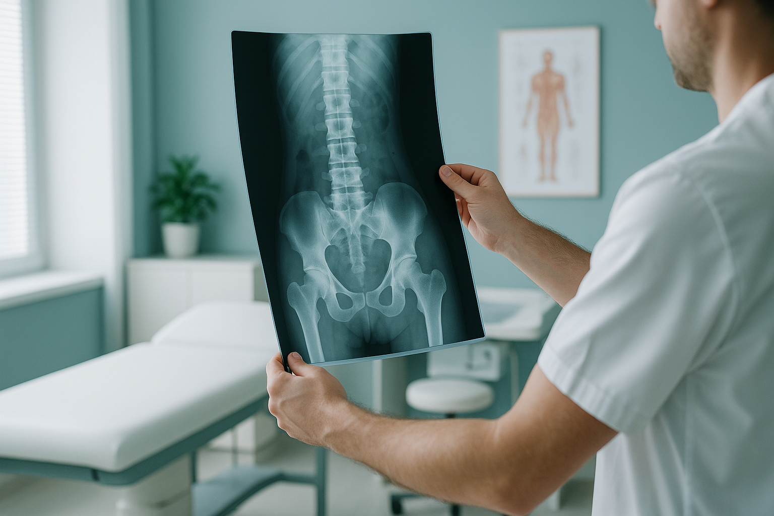
Full spine x-ray stitching means combining two or more separate x-ray exposures into one long image. A standard x-ray detector covers roughly a third of the spine at a time. So the machine takes overlapping shots of the upper, middle and lower sections. Software then lines up the overlapping areas and merges them into what looks like one image.
This method is sometimes called long film radiography. It has been used for decades. Before single-pass technology existed, it was the only practical way to see the whole spine on one image. Most hospital radiology departments still use it today.
Why do full spine x-rays need stitching?
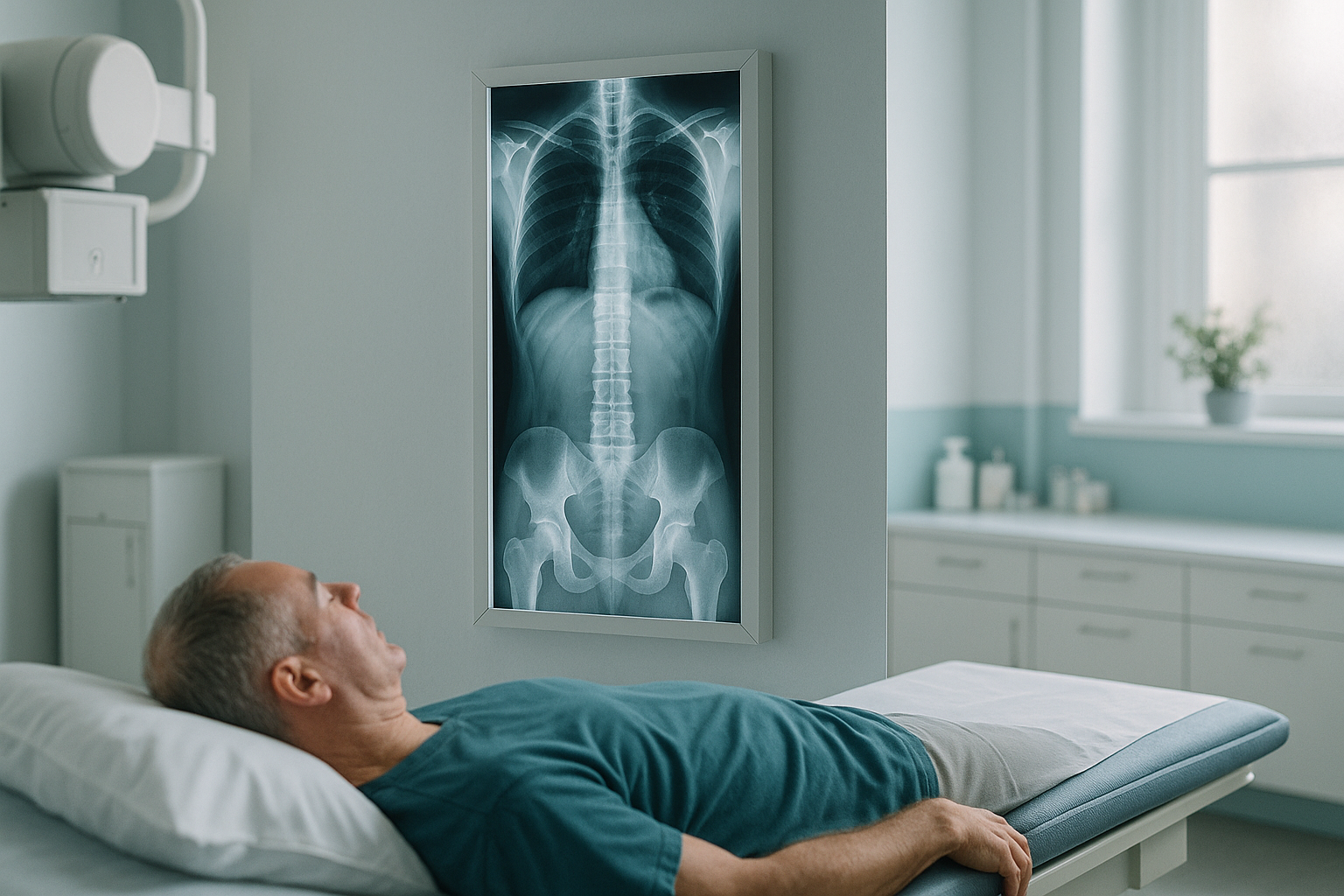
They need stitching because no standard x-ray detector is tall enough to capture the full spine in one exposure. In an adult, the spine from the base of the skull to the pelvis is usually longer than even the largest flat panel detector. To get a full-length view, the machine must take separate shots and combine them.
Standard panels cover only part of the spine in each shot.
Each section needs its own exposure, which adds time and radiation.
Software must find matching anatomy in the overlapping areas to join the frames.
Each extra exposure also adds to the total radiation dose. If images must be repeated because the stitch failed, the dose goes up further.
What errors can happen with stitched spine x-rays?
Several problems can appear when x-ray frames are joined. Some are easy to see on the image. Others cannot be seen by eye but still affect the measurements.
A visible line or step at the join between two frames, where the anatomy does not line up perfectly.
Even a slight shift, sway or deep breath between exposures changes the position of the spine from one frame to the next.
Poorly aligned stitching can change how the curve of the spine looks, which leads to inaccurate alignment values.
When the stitch clearly fails, the whole study may need repeating, which doubles the radiation exposure.
Why measurement distortion matters
Surgeons and physiotherapists rely on precise spine measurements when planning treatment. If a stitched image shifts the apparent alignment by even a small amount, it can change the treatment advice. For example, a curve that looks borderline on a stitched image might really be larger or smaller than it appears. In the same way, the sagittal vertical axis, which describes how your head lines up over your pelvis from the side, can be thrown off by a stitching error.
The specialist reading the image may not always know that a measurement is unreliable, because the seam is not always visible. This affects how confident the report can be, and in turn the decisions made about your care.
How does single-pass imaging avoid stitching problems?
Single-pass imaging captures the whole body in one continuous sweep, so there is nothing to stitch. The scanner moves from head to feet in one motion while you stand naturally. The result is one unbroken image with no seams, no overlap areas and no risk of movement between frames.
Several exposures. Two or three separate shots taken one after another.
Movement risk. You must hold still for each exposure. Any shift between frames distorts the final image.
Software joining. Overlap areas are matched digitally. If the anatomy moves, the software produces a seam artefact.
Higher total dose. Several exposures, plus possible repeats, add up.
One continuous sweep. The system scans from top to bottom in one pass while you stand upright.
No joins. There is no stitching step. No overlap areas. No seam artefacts.
True weight-bearing alignment. Measurements come from one image, captured in one moment, so they reflect your real standing posture.
Lower radiation. About 90% less radiation than a standard x-ray, around ten times less.
This is the approach used by ScanAlign, unstitched full-body imaging. Because the image is captured in one pass while you stand, the alignment values reflect your real posture, not a combined image that stitching may have slightly distorted.
Worried about the accuracy of an earlier stitched x-ray? A single-pass standing scan removes the guesswork.
Book a free video consultationWhen MRI is the better choice. EOS and x-ray both show bones, joints and alignment. For soft tissue, discs or nerves, MRI is usually the better tool. If the specialist treating you suspects a disc bulge or nerve compression, they may recommend MRI alongside or instead of an alignment scan.
Before you start treatment, or change the one you are on, it is worth getting a detailed diagnosis. Knowing whether the cause sits in the pelvis, the legs or the spine changes what will actually help.
Frequently asked questions about x-ray stitching
It usually shows as a faint horizontal line or a slight step where one frame meets the next. Sometimes the anatomy above and below the line does not quite match. It may be so subtle that only a trained eye spots it.
It can. If the stitch shifts the apparent position of the vertebrae, the measured curve or the sagittal vertical axis may be inaccurate. This could lead to slightly different medical advice from the advice you would get from an unstitched image.
They are fairly common, though how serious they are varies. Even well-calibrated systems can produce minor misalignment if the patient shifts between exposures. Published research has noted measurable differences in alignment values between stitched and single-pass images.
No. It feels much the same. You stand still while the machine takes each shot. The challenge is that holding perfectly still for several exposures over a longer time is harder than standing for one quick sweep.
A single-pass image removes stitching as a variable completely, so there is no seam artefact and no risk of the patient moving between frames. This generally makes measurements more reliable, especially for alignment values that cover the full length of the spine.
No. ScanAlign is a self-referral clinic. You can book directly without a letter from your doctor or a consultant. Your scan is reviewed by a specialist radiographer and reported by a consultant radiologist.
An EOS scan uses about 90% less radiation than a standard x-ray, around ten times less. Because there is only one pass, you are never exposed to the repeat doses that can happen when a stitched image has to be taken again.
The price for patients travelling from abroad is confirmed during the free video consultation.
Yes. If your first scan has a stitching artefact and your follow-up scan does not, the measurements may not be directly comparable. This makes it harder to tell whether a curve or alignment shift has truly changed over time.
Some NHS hospitals in the UK do have EOS systems, but availability varies. ScanAlign at 19 Harley Street, London is a private, self-pay clinic offering standing EOS imaging. You do not need a referral to book.
Get an unstitched, full-body standing image
One continuous sweep. No joins. No guesswork. See your alignment as it truly is.
Book a free video consultationSources
- Wybier M, Bossard P. Musculoskeletal imaging in progress: the EOS imaging system. Joint Bone Spine. 2013;80(3):238-243.
- Illés T, Somoskeöy S. The EOS imaging system and its uses in daily orthopaedic practice. International Orthopaedics. 2012;36(7):1405-1411.
- The Royal College of Radiologists. Standards for the reporting and interpretation of imaging investigations. Second edition, 2018.
Written by Abbas Dhami (Specialist Diagnostic Radiographer).
This page is general information and not a diagnosis.
ScanAlign is the trading name of CoreMed Solutions Ltd and operates under The Harley Street Hospital's CQC licence.