
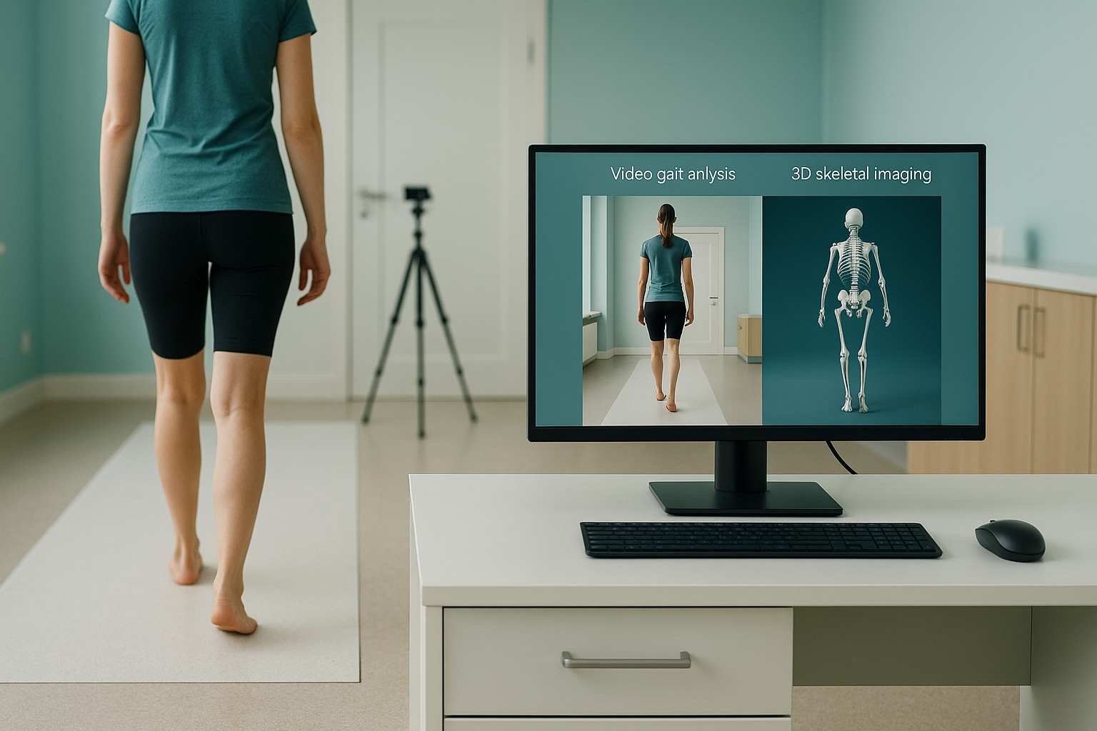
Video Gait Analysis or 3D Skeletal Imaging: What Is the Difference?
Video gait analysis records how you move, usually on a treadmill. It shows muscle timing, stride pattern and how your foot strikes the ground. 3D skeletal imaging, such as a standing EOS scan, captures the real shape and alignment of your bones under load. One watches movement. The other shows the structure behind it. They answer different questions, and they often work best together.
Key points
- Video gait analysis shows how your body moves in real time. But it films the surface of the skin, not the skeleton.
- 3D skeletal imaging (EOS) captures exact bone shape and joint alignment while you stand with your full weight on your legs.
- Skin markers can shift during movement. This limits how precise video measurements can be.
- The two tests complement each other. Movement data explains the "how". Skeletal data explains the "why".
- Neither one replaces MRI when detail of soft tissue, discs or nerves is needed.
What is video gait analysis and what does it show?
Video gait analysis is a movement assessment. A camera, or sometimes several cameras, records you walking or running, often on a treadmill. A clinician or software then reviews the footage frame by frame. It shows stride length, cadence, foot strike pattern, knee tracking, and how your pelvis, trunk and arms move with each step.
Many running shops, physiotherapy clinics and sports medicine centres offer treadmill gait analysis. It is useful for spotting compensations, timing faults and imbalance between the two sides of your movement. Some labs add pressure plates or force sensors to measure ground reaction forces.
What is 3D skeletal imaging?
3D skeletal imaging creates a precise picture of your bones and joints that can be measured. An EOS scan, for example, takes a full-length image from head to foot while you stand naturally. Because you are upright and bearing your own weight, the image shows how gravity and your body weight affect your skeleton. A specialist can then measure bone length, joint angles, rotation and overall lower limb alignment from the 3D reconstruction.
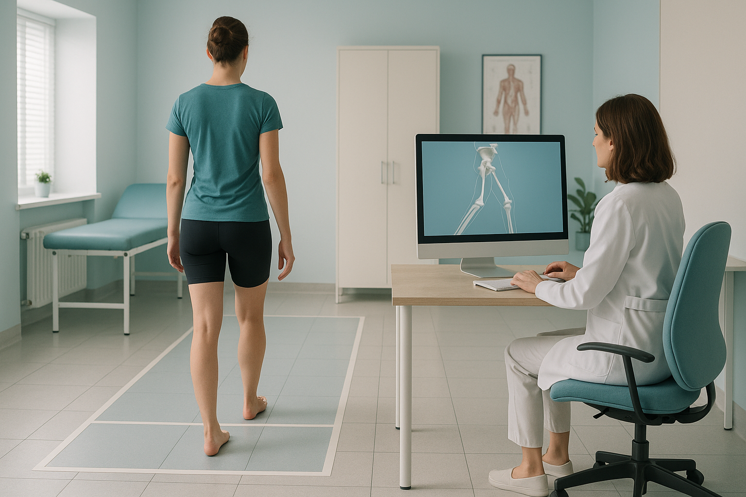
How does video gait analysis compare with 3D skeletal imaging?
The two tests measure very different things. The table below sums up the main differences.
| Feature | Video gait analysis | 3D skeletal imaging (EOS) |
|---|---|---|
| What it captures | Surface movement patterns while walking or running | Bone shape, length, rotation and joint alignment |
| Position | Moving on a treadmill or walkway | Standing still, full weight on the legs |
| Static or dynamic | Dynamic (in motion) | Static (standing alignment under load) |
| Markers | Skin markers or camera tracking without markers | No markers needed. The bones are imaged directly. |
| How it measures | Joint centres estimated from the skin surface | True bone shape from a calibrated radiograph |
| Radiation | None | About 90% less radiation than a standard X-ray, around ten times less |
| Soft tissue detail | None (outside view only) | Bones and joints only. MRI is better for soft tissue. |
| Best for | Finding movement faults, running technique, choosing shoes | Diagnosing structural causes such as leg-length difference, torsion and joint malalignment |
Why can video gait analysis miss structural problems?
Video gait analysis is valuable, but it has limits that are worth understanding. It records what happens on the outside of the body. It cannot see bones, joint surfaces or internal rotation.
These limits do not make video gait analysis wrong. They simply mean it answers a different question. It tells you how you move. It does not tell you why your skeleton is shaped the way it is.
How does standing EOS imaging add to a gait assessment?
An EOS scan images your whole skeleton from head to foot while you stand. Because your muscles, joints and gravity are all at work, the image shows your real alignment under load. Lying-down scans relax the body and can hide problems that only show up when your body is upright and carrying its weight.
Your body is a connected chain. A twist in the shinbone can change how the knee tracks. That shifts the hip, which tilts the pelvis. If you only look at one joint, you may see the compensation but miss the cause. A full-length standing image captures the whole chain in one view. It gives clinicians EOS compared with X-ray, CT and MRI context they cannot get from traditional imaging alone.
Looking at your postural sway and static alignment together with your movement data gives the fullest picture. The gait video shows the movement fault. The skeletal image shows the structural reason behind it.
Want to see what your skeleton looks like under load? At ScanAlign, standing EOS imaging in London, a standing whole-body scan takes about 20 seconds.
Book a free video consultationWhen should you choose gait analysis, skeletal imaging, or both?
It depends on your question. If you want to improve your running form or check how your shoes fit, a video gait assessment may be all you need. If you have a persistent structural concern, such as a leg that feels longer, a knee that keeps giving way, or hip pain that keeps coming back after years of physiotherapy, skeletal imaging can show what is happening at bone level.
- Want to improve your running or walking technique
- Need help choosing the right footwear
- Want to see how your muscles fire during movement
- Have a sports coach or physiotherapist guiding your training
- Have ongoing pain that has not responded to treatment
- Suspect a leg-length difference or a bone rotation problem
- Need alignment measurements before surgery
- Want to understand why a gait pattern is abnormal, not just that it is
Many people benefit from both. A physiotherapist reviews the gait data, a radiologist reviews the skeletal data, and together they build a clearer picture. You can book a skeletal alignment scan without a referral from your doctor.
Before you start a treatment, or change the one you are on, it is worth getting a detailed diagnosis. Knowing whether the cause sits in your pelvis, your legs or your spine changes what will actually help.
When MRI is the better choice: EOS shows bones, joints and alignment. For soft tissue, discs or nerves, MRI is usually the better tool. If your clinician suspects a ligament tear, cartilage damage or a compressed nerve, an MRI is likely more suitable than either gait analysis or skeletal imaging.
Frequently asked questions
Video gait analysis is designed to assess movement patterns, not bone structure. It can point out abnormal motion, but it cannot diagnose conditions like bone torsion, leg-length differences or joint deformity. For those, you need imaging that shows the skeleton directly.
It usually measures stride length, cadence, foot strike angle, knee tracking and pelvic movement while you walk or run. Some systems also record ground reaction forces through a pressure plate under the treadmill belt.
An EOS scan images your whole body from head to foot in a single upright pass with about 90% less radiation than a standard X-ray, around ten times less. It also produces a 3D model of your skeleton, which a standard X-ray cannot do.
No. ScanAlign is a self-pay clinic, and you can book directly without a referral from your doctor or a specialist. Your first step is a free video consultation to check the scan is right for you.
Yes. The two tests complement each other. Many clinicians recommend combining movement data from a gait lab with structural data from a standing skeletal scan for the most complete picture.
Skin markers are small reflective dots placed on landmarks like the knee or ankle. As the body moves, the skin and the soft tissue under it shift. This moves the marker away from the true position of the bone. This is called soft tissue artefact, and it can cause measurement errors in video-based analysis.
Gait analysis costs vary widely depending on the clinic and the technology used. For patients travelling from abroad, the price of an EOS scan at ScanAlign is confirmed during the free video consultation, and you can read more on the FAQs and cost page.
An EOS scan uses about 90% less radiation than a standard X-ray, around ten times less. The dose is very low. Your radiographer will explain the details during your consultation.
It may show that your knee collapses inward during your stride, which is useful. But it cannot tell you whether that movement is caused by muscle weakness, bone rotation or a joint alignment problem. If the problem persists, imaging of the skeleton may help find the structural cause.
Sudden leg weakness, numbness or loss of bladder or bowel control can be a sign of a serious condition that needs urgent attention. Please contact your doctor straight away or go to the nearest emergency department, rather than booking an imaging appointment.
See the structure behind the movement
A free video consultation helps you find out whether a standing skeletal scan is the right next step for your concern.
Book a free video consultationWritten by Abbas Dhami (Specialist Diagnostic Radiographer)
Sources
- Leardini A, Chiari L, Della Croce U, Cappozzo A. Human movement analysis using stereophotogrammetry. Part 3: Soft tissue artifact assessment and compensation. Gait & Posture. 2005;21(2):212-225.
- Illés T, Somoskeöy S. The EOS imaging system and its uses in daily orthopaedic practice. International Orthopaedics (SICOT). 2012;36(7):1375-1386.
- Royal College of Radiologists. iRefer: Making the best use of clinical radiology. Available at: irefer.org.uk.
This page is general information and not a diagnosis.
ScanAlign is the trading name of CoreMed Solutions Ltd and operates under The Harley Street Hospital's CQC licence.