
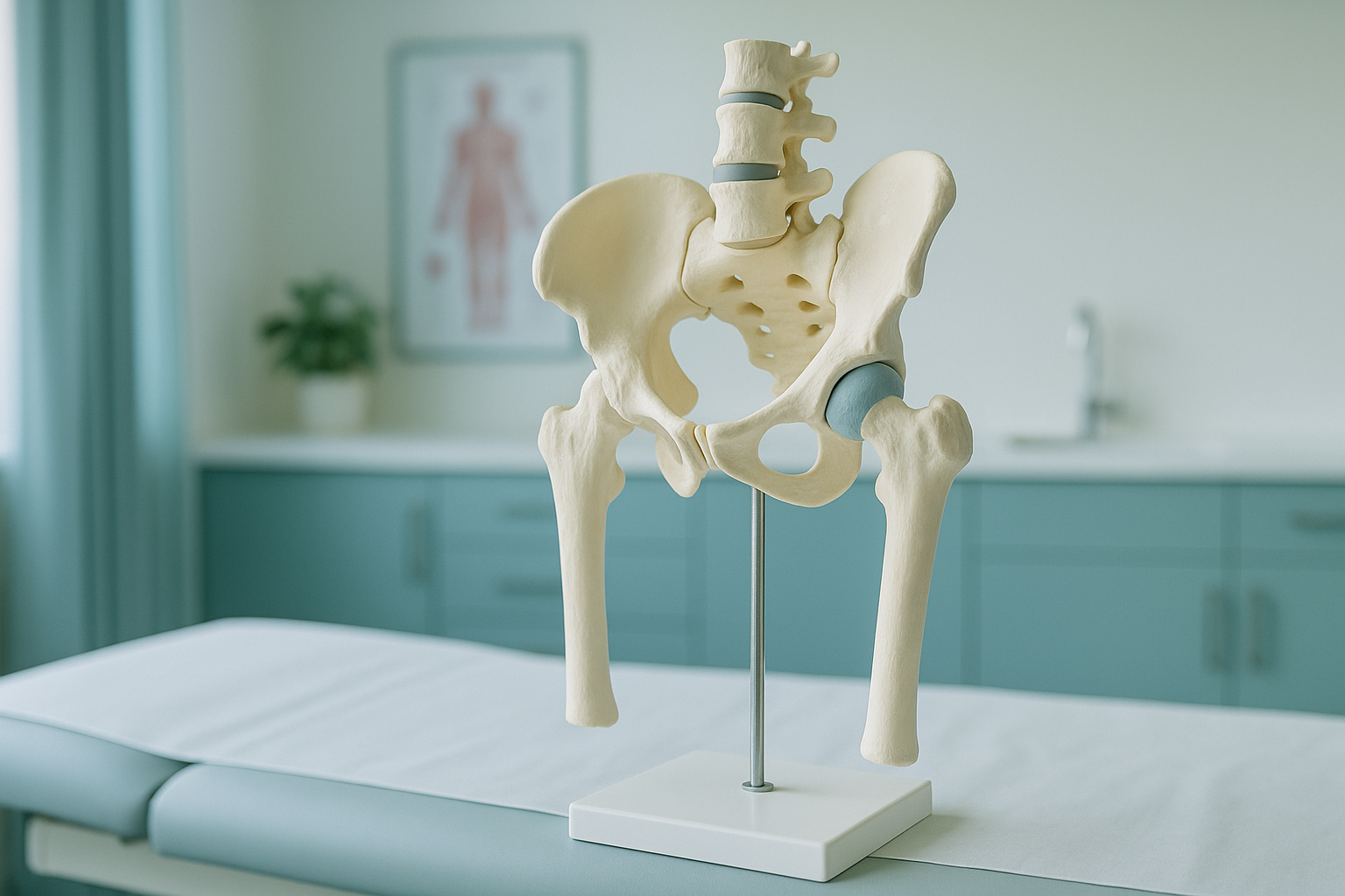
Hip and Pelvis Anatomy: How Does the Hip Joint Actually Work?
The hip joint is a ball and socket joint. The rounded head of the thigh bone (femur) sits inside a cup-shaped socket in the pelvis (acetabulum). It is one of the strongest and most stable joints in the body. It carries your full body weight when you stand and walk. Its alignment affects everything from your lower back to your knees.
Key points
- The hip is a ball and socket joint built for stability and a wide range of movement.
- The pelvis is made of three fused bones (ilium, ischium, pubis). It passes load between your spine and your legs.
- Muscles, ligaments and the labrum all work together to keep the joint stable.
- Scans done lying down can miss alignment problems that only appear when you are standing and bearing weight.
- A standing EOS scan shows how bones, joints and soft tissue work together under real-life load.
Knowing how your hip is built helps you make sense of pain, stiffness or clicking. It also shows why a problem in one area often causes symptoms somewhere else. This page explains each part of the hip joint and pelvis in plain language. It also explains why standing imaging matters for an accurate picture.
Which bones make up the hip joint and pelvis?
The hip joint forms where two bones meet: the femur (thigh bone) and the pelvis. The rounded top of the femur is called the femoral head. It fits into a deep cup in the pelvis called the acetabulum. This ball and socket design gives the hip both strength and flexibility.
The pelvis itself is made of three bones that fuse together during the teenage years:
- Ilium: the large, wing-shaped bone you feel at your waist.
- Ischium: the lower part you sit on.
- Pubis: the front section, where the left and right sides meet at the pubic symphysis.
At the back, the pelvis joins the base of the spine through the sacroiliac (SI) joints. These joints pass load between your upper body and your legs. When the SI joint is not working properly, it can cause pain that feels as if it is coming from the hip or lower back. You can read more about SI joint dysfunction and how it links to hip and back pain.
The angle at which the femoral head sits in the socket also matters. If the ball points too far forward or too far back, it changes how the joint moves and where stress falls. This is called hip anteversion and retroversion. It can only be measured accurately with the right imaging.
What do the labrum and ligaments do in the hip?
The labrum is a ring of tough, flexible cartilage that lines the rim of the acetabulum. It deepens the socket, helps seal the joint fluid inside, and improves the grip between the ball and the socket. A damaged labrum is called a hip labral tear. It can cause clicking, catching or a deep ache in the groin.
The hip capsule is a tough, fibrous sleeve that wraps around the whole joint. Three main hip ligaments make it stronger. The strongest is the iliofemoral ligament. It runs across the front of the hip and stops you tipping backwards when you stand upright. These ligaments are under real tension only when you are bearing weight. This is one reason standing imaging gives a more accurate picture than imaging done lying down.
Which hip muscles control movement and stability?
More than twenty muscles cross the hip joint. They work in groups to move and steady the joint during every step, squat and turn. Here are the main ones.
These hip muscles do not work alone. They are part of a connected chain that runs from your feet up through your pelvis and spine. For example, a tight iliopsoas can tilt the pelvis, change the curve of the lower back and put extra load on the opposite knee. This is why pain often shows up in a different place from its true cause.
What range of movement does the hip joint allow?
The ball and socket design of the hip allows movement in all directions: forward (flexion), backward (extension), out to the side (abduction), across the body (adduction) and turning inward and outward (rotation). This wide range of movement is what lets you walk, run, sit cross-legged and climb stairs.
The price of this freedom is complexity. So many parts work together that a small change in one of them can alter how load passes through the whole pelvis. That change might be the angle of the femoral head, the depth of the socket or the tension of a single ligament. Over time, the body compensates. You may not feel it in the hip at all. Instead, the first symptom might be a sore lower back, a stiff SI joint or a nagging knee.
If you have persistent hip, back or knee pain that has not been explained, understanding your alignment under load is the logical next step.
Book a free video consultationWhy does standing imaging show hip problems that lying-down scans miss?
When you lie down for a standard X-ray, CT scan or MRI, gravity is taken out of the picture. Your muscles relax, your pelvis settles into a neutral position, and alignment problems can disappear. This is why a scan may come back "normal" even when you are in pain every time you stand or walk.
A standing EOS scan captures your hip joint, pelvis and both legs in full under your real body weight. It shows how load passes through the SI joints, whether the pelvis is tilted or rotated, and how the femoral head sits in the socket when gravity is acting on it. This is the picture your surgeon or physiotherapist needs to plan treatment accurately.
Imaging done lying down removes the effect of gravity and muscle tension. A pelvic tilt, a difference in leg length or a shift in alignment may flatten out completely. The images look normal, but your symptoms continue, because the real problem only appears when you are upright and bearing weight.
ScanAlign standing EOS imaging uses about 90% less radiation than a standard X-ray, around ten times less. A single scan captures your full skeleton from head to toe while you stand naturally. This gives a complete picture of how your bones and joints relate to each other under load.
A note on MRI. EOS is excellent at showing bones, joints and alignment under load. For soft-tissue problems such as disc injuries, nerve compression or muscle tears, MRI is usually the better tool. The two types of imaging work well together.
Before you start a treatment, or change the one you are on, it is worth getting a detailed diagnosis first. Knowing whether the cause sits in your pelvis, your legs or your spine changes what will actually help.
Frequently asked questions about hip and pelvis anatomy
The hip is a ball and socket joint. The rounded femoral head (ball) fits into the acetabulum (socket) of the pelvis. This design allows movement in all directions while keeping the joint very stable.
The pelvis is made of the ilium (the broad upper wing), the ischium (the lower part you sit on) and the pubis (the front). These three bones fuse into one solid structure during the teenage years.
The labrum is a ring of cartilage around the rim of the hip socket. It deepens the socket, helps spread pressure evenly, and seals in joint fluid to keep the cartilage surfaces lubricated.
Yes. The body is a connected chain. A change in hip alignment, muscle tightness or pelvic tilt can shift load onto the lower back, SI joints or knees. Pain often appears away from the original cause.
When you lie down, gravity is removed and alignment problems can disappear. A standing scan captures your hip under your real body weight. It shows how load passes through the pelvis and whether the joint is positioned correctly when you are upright.
EOS uses about 90% less radiation than a standard X-ray, around ten times less. It is one of the lowest-dose imaging methods available for full skeletal assessment.
No. ScanAlign is a private, self-pay clinic. You can refer yourself directly. The first step is a free video consultation to discuss whether a standing scan is right for you.
The hip joint is where the thigh bone meets the pelvis. The sacroiliac (SI) joint is where the pelvis meets the base of the spine. They are separate joints, but problems in one often affect the other, because they share load when you stand and move.
For patients travelling from abroad, the price is confirmed during the free video consultation, which you can arrange from the book a standing hip and pelvis scan page.
EOS is designed for bone and joint alignment. For soft-tissue injuries such as labral tears, cartilage damage or muscle problems, MRI is usually the better tool. The two types of imaging work well together.
See how your hip and pelvis work under load
A free video consultation, from wherever you are, will help you understand whether a standing EOS scan at 19 Harley Street, London is the right next step for your symptoms.
Book a free video consultationWritten by Abbas Dhami (Specialist Diagnostic Radiographer)
Sources
- Gray's Anatomy: The Anatomical Basis of Clinical Practice, 42nd edition. Elsevier, 2020. Chapters on the pelvis and lower limb.
- Byrne DP, Mulhall KJ, Baker JF. "Anatomy & Biomechanics of the Hip." The Open Sports Medicine Journal, 2010; 4:51-57.
- NHS. "Hip pain in adults." nhs.uk/conditions/hip-pain. Accessed 2024.
This page is general information and not a diagnosis.
ScanAlign is the trading name of CoreMed Solutions Ltd and operates under The Harley Street Hospital's CQC licence.