
Osteomalacia: What Causes Soft Bones in Adults and How Is It Treated?
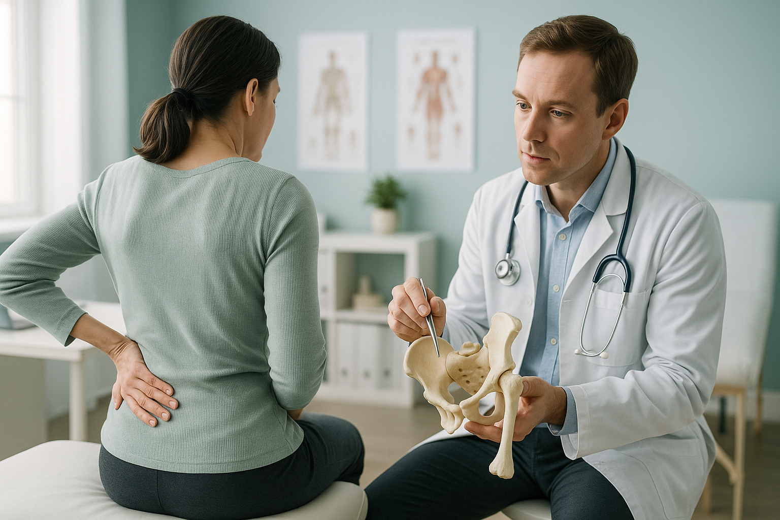
Osteomalacia is a condition where bones become soft because they do not have enough minerals. This is usually due to a lack of vitamin D. The main treatment is to correct that deficiency with supplements and diet changes, and sometimes by treating an underlying cause. Imaging helps assess how far bone softening has affected your skeleton and posture.
Key points
- Osteomalacia means your bones are not mineralising properly. This makes them weak and painful.
- In adults, the most common cause is not getting enough vitamin D.
- It is different from osteoporosis, although both affect bone strength.
- Treatment usually centres on vitamin D and calcium supplements, guided by your doctor.
- Standing whole-body imaging can reveal alignment changes and stress areas that lying-down scans may miss.
What is osteomalacia in adults?
Osteomalacia is a bone disorder where newly formed bone tissue fails to mineralise. This leaves it softer than normal. Healthy bones need a steady supply of calcium and phosphate. These are laid down into a protein framework to make hard, strong bone. When vitamin D levels are too low, or another condition disrupts mineral absorption, this process stalls. The bone structure is still there, but it stays rubbery rather than rigid.
In children, the same problem is called rickets. In adults, it is called osteomalacia. The condition develops slowly, so many people put up with vague aches for months or years before they get a diagnosis.
What are the symptoms of osteomalacia?
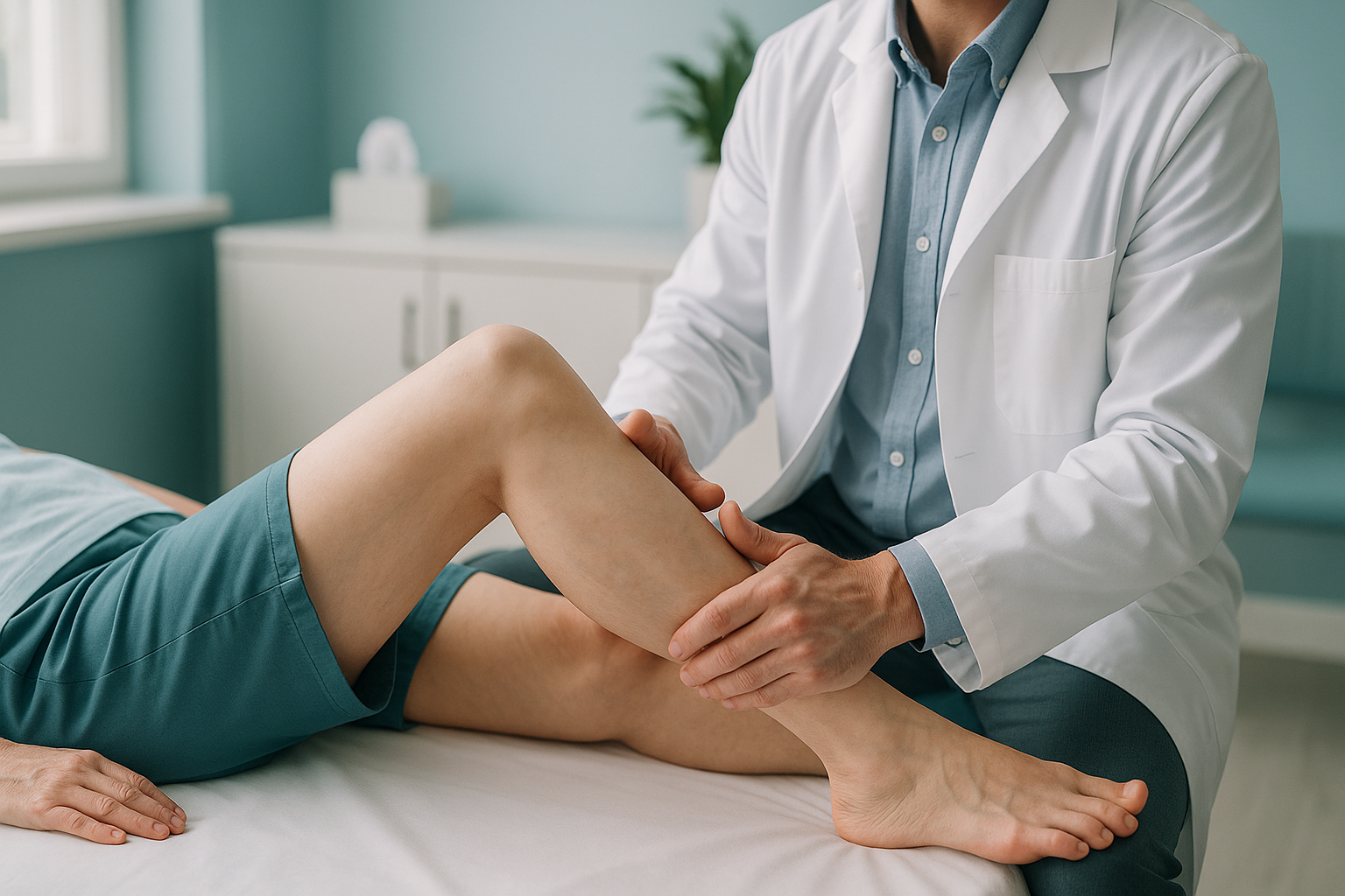
The most common symptoms of osteomalacia are a deep, aching bone pain and muscle weakness. These are felt especially in the hips, pelvis, lower back and legs. Pain tends to be worse when you put weight through the affected bones, so standing and walking often feel harder than they should.
A dull, persistent ache in the hips, pelvis, lower back or legs that gets worse with activity.
Difficulty climbing stairs, getting up from a chair or walking longer distances.
A side-to-side way of walking caused by weak hip and thigh muscles.
These symptoms overlap with many other conditions. Because of this, osteomalacia is sometimes mistaken for fibromyalgia, arthritis or general tiredness. If bone pain has lasted for weeks without a clear explanation, it is worth asking your doctor about a vitamin D blood test.
What causes osteomalacia?
The most common cause of osteomalacia is a lack of vitamin D. Your body needs vitamin D to absorb calcium from food. Without it, not enough calcium reaches the bones, and mineralisation stalls. Several factors can lead to low vitamin D or poor mineral absorption.
Skin makes vitamin D from sunlight. Spending most of the day indoors, wearing covering clothing or living at northern latitudes all reduce how much is made.
A diet low in vitamin D and calcium, particularly one without oily fish, eggs or fortified foods.
Conditions such as coeliac disease or Crohn's disease can stop the gut from absorbing vitamin D or calcium properly.
These organs turn vitamin D into its active form. If they are not working well, the active form may be too low.
Some medicines, including some anti-seizure drugs, can also interfere with how the body uses vitamin D. Your doctor will want to find the underlying cause before starting treatment, so that the right approach is used.
How is osteomalacia different from osteoporosis?
Both conditions weaken bones, but in different ways. Osteomalacia is a mineralisation problem: there is enough bone structure, but it is not hardened properly. Osteoporosis is a density problem: the overall amount of bone tissue goes down, making bones thin and brittle.
What happens: New bone fails to mineralise. The bone framework is there but stays soft.
Main cause: Lack of vitamin D or problems absorbing minerals.
Typical symptom: Deep, aching bone pain, muscle weakness.
Reversible? Often yes, once the deficiency is corrected.
What happens: Bone tissue is lost faster than it is replaced, which reduces bone density.
Main cause: Ageing, hormonal changes (especially after menopause), genetics.
Typical symptom: Often silent until a fracture happens.
Reversible? Bone density can be improved but usually not fully restored.
It is possible to have both conditions at the same time. If you have unexplained bone pain and risk factors for either, a proper assessment matters.
How is osteomalacia diagnosed?
Diagnosis usually starts with a blood test. Your doctor will check your levels of vitamin D, calcium, phosphate and alkaline phosphatase (an enzyme that rises when bones are not mineralising properly). X-rays can show typical changes, such as thinning of the bone cortex or small stress fractures called Looser zones. In some cases a bone biopsy is used, though this is uncommon.
Standard X-rays are taken lying down. This means they only show one area at a time and do not show how softened bones cope with your body weight. This is where standing, whole-body imaging adds real value.
Why does standing imaging matter for osteomalacia?
When you lie down for a normal X-ray or MRI, gravity is taken out of the picture. Your muscles relax and your joints decompress. Any bowing or shift in alignment caused by softened bone is partly hidden. Standing whole-body EOS imaging captures your skeleton while it carries your real weight, from your ankles to your skull, in a single scan.
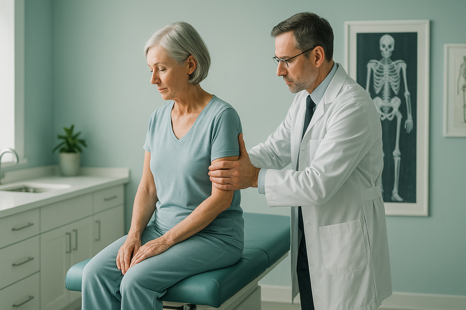
Your body is a connected chain. When one bone softens or bows under load, the joints above and below compensate. Pain may appear in your knees, hips or lower back rather than at the weakened bone itself. A whole-body standing scan maps this whole chain, so your treating clinician can see the full picture.
EOS technology uses about 90% less radiation than a standard X-ray, around ten times less. That makes it a practical option when a full-length view of your skeleton is needed.
Before you start treatment, or change the one you are on, it is worth getting a detailed diagnosis. Knowing whether the cause sits in the pelvis, the legs or the spine changes what will actually help.
Would you like to see how your bones and alignment look when you are standing?
Book a free video consultationWhen is MRI more useful? EOS imaging shows bones, joints and alignment very well. For soft-tissue detail, such as muscles, tendons or nerves, MRI is usually the better tool. If your doctor suspects a soft-tissue cause alongside osteomalacia, they may recommend MRI as well.
What does osteomalacia treatment involve?
Treatment focuses on correcting the underlying deficiency so that your bones can start mineralising again. In most cases, this means vitamin D and calcium supplements prescribed by your doctor or specialist. The exact dose and how long you take it depend on how severe the deficiency is and what caused it.
- Vitamin D supplements are the cornerstone of treatment. Your doctor will choose the right form and dose based on your blood test results.
- Diet changes may be recommended, such as eating more oily fish, eggs, fortified cereals and dairy products.
- Treating the underlying cause is important. If gut malabsorption or a kidney condition is involved, dealing with that condition helps vitamin D levels recover.
- Monitoring with follow-up blood tests makes sure levels are improving and bones are responding.
ScanAlign is a diagnostic imaging clinic, not a treatment provider. We mention treatments here to give you a clear picture of the condition, but any treatment decisions should be made with your doctor or specialist. What we can do is provide detailed standing alignment images that help your clinician plan the right care.
Important: if you have sudden severe bone pain, cannot bear weight, or notice a new deformity, please get urgent medical help from your doctor or your nearest emergency department rather than booking a scan.
Frequently asked questions about osteomalacia
No. Osteomalacia means bone is not mineralising properly, which leaves it soft. Osteoporosis means overall bone density is reduced, which makes bones thin and brittle. Both weaken bones, but they have different causes and different treatments.
In many cases, yes. When the vitamin D deficiency is corrected and any underlying cause is treated, bones can start to mineralise normally again. How quickly things improve depends on how severe the deficiency was and how long it lasted.
Most people describe a deep, dull ache in the bones, particularly around the hips, pelvis, lower back and thighs. The pain tends to be worse when you stand, walk or put weight through the affected area. It often builds up slowly over weeks or months.
Vitamin D helps your body absorb calcium from food. Without enough vitamin D, not enough calcium reaches your bones for proper mineralisation. The result is softer bone tissue, which aches under normal daily loads.
No. ScanAlign is a private, self-pay clinic. You can refer yourself. If you would like to discuss whether a standing alignment scan is right for you, you can start with a free video consultation.
Yes. EOS uses about 90% less radiation than a standard X-ray, around ten times less. This lower dose makes it a practical choice when full-body or repeat imaging is needed. You can read more on the FAQs and safety page.
For patients travelling from abroad, the price is confirmed during the free video consultation. You can find more answers on the ScanAlign FAQs page.
Yes. Softened bone is more vulnerable to stress fractures and can break from fairly minor impacts. This is one reason early diagnosis matters, so treatment can start before fractures happen.
When you lie down, gravity no longer acts on your skeleton. Muscles relax, joints decompress, and any bowing or tilt caused by softened bone can partly straighten out. Standing imaging captures what your body actually does under your own weight. This gives a more accurate picture of alignment.
Your images are reviewed by a specialist reporting team. You receive a detailed report that you can share with your doctor, consultant or physiotherapist to guide your next steps. ScanAlign provides the diagnostic imaging, not the treatment itself.
See how your bones and alignment look when you are standing
A free video consultation with our team can help you decide whether a standing whole-body scan in London is the right next step.
Book a free video consultationRelated pages
Sources
- NHS. "Vitamin D." nhs.uk. Overview of vitamin D deficiency, causes and treatment guidance.
- Bhan A, Rao AD, Rao DS. "Osteomalacia as a result of vitamin D deficiency." Endocrinology and Metabolism Clinics of North America, 2010;39(2):321-331.
- National Institute for Health and Care Excellence (NICE). "Vitamin D deficiency in adults: treatment and prevention." Clinical Knowledge Summary.
Written by Abbas Dhami (Specialist Diagnostic Radiographer)
This page is general information and not a diagnosis.
ScanAlign is the trading name of CoreMed Solutions Ltd and operates under The Harley Street Hospital's CQC licence.