
What Are Pelvic Incidence, Sacral Slope and Pelvic Tilt?
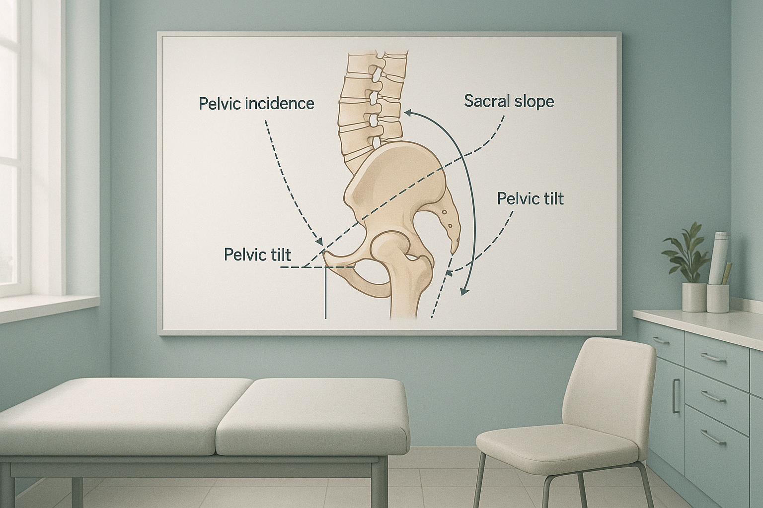
Pelvic incidence, sacral slope and pelvic tilt are three measurements taken from your pelvis and the base of your spine. Together they describe the shape and angle of your pelvis. This directly controls the curves of your spine. They can only be measured accurately on an image of your whole body taken while you stand and bear your own weight.
Key points
- Pelvic incidence (PI) is a fixed value set by the shape of your bones. It does not change after you stop growing.
- Sacral slope and pelvic tilt depend on your position. They shift when you stand, sit or lean.
- PI always equals sacral slope plus pelvic tilt. If one changes, the other must make up for it.
- A mismatch between PI and lordosis is one of the most common causes of sagittal balance problems.
- These values can only be measured properly on a standing, full-body X-ray.
What do pelvic incidence, sacral slope and pelvic tilt each measure?
Each of the three spinopelvic parameters measures a different part of the shape of your pelvis. They work as a linked set of three: change one, and the others must adjust.
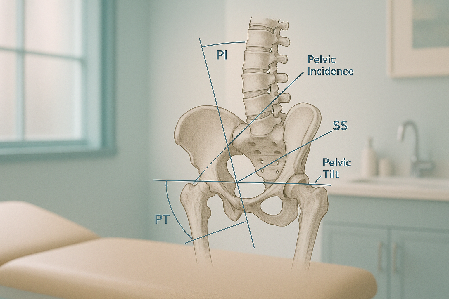
A fixed value, set by your anatomy, that describes the overall shape and angle of your pelvis. It comes from your bone structure and does not change with posture. A higher PI means your body needs a deeper curve in the lower back to stay balanced.
The angle at which the top surface of your sacrum (the triangular bone at the base of the spine) tilts forward. A steeper sacral slope usually goes together with a deeper lumbar curve.
How far the pelvis has rotated backward (retroversion) or forward in relation to the hip joints. Your body adjusts pelvic tilt to make up for it when the spine loses its natural curves.
PI is fixed and equals sacral slope plus pelvic tilt. So your body can only shift the balance between the other two. If the sacral slope drops (the sacrum becomes flatter), the pelvic tilt must increase. This is exactly what happens when someone develops flatback syndrome: the pelvis tilts backward to keep the head over the feet.
Why do spinopelvic parameters matter for back pain?
Spinopelvic parameters matter because they decide the size and shape of every curve above the pelvis. Your spine is a chain. The pelvis is its foundation. If the foundation is at a steep angle (high PI), the lumbar spine must curve more to make up for it. If the curves do not match the pelvis, the whole chain falls out of sagittal balance. Muscles, joints and discs then take extra strain.
Many people with persistent low back pain, hip pain or difficulty standing upright have a mismatch between these values. The pain they feel in the lower back, hips or even the knees is often the result of the body making up for this. It is not a problem at the painful spot itself. Understanding the pelvis is often the key to understanding the pain.
What is a PI-LL mismatch and why does it cause problems?
A PI-LL mismatch happens when pelvic incidence and lumbar lordosis are not well matched. In a balanced spine, the depth of the lumbar curve should closely follow the pelvic incidence value. When lordosis is much less than PI, the body's centre of gravity shifts forward.
To keep your head over your feet, your body makes up for it. It may tilt the pelvis backward (increased pelvic tilt), flatten the mid-back, bend the knees, or tighten the muscles that straighten the hips. You might feel this as:
- Difficulty standing upright for more than a few minutes
- A constant feeling of falling forward
- Aching in the lower back, buttocks or backs of the thighs
- Tiredness after walking short distances
PI-LL mismatch is one of the most studied measures in planning spine surgery. Surgeons rely on this link to decide how much correction is needed and where. Without accurate spinopelvic numbers from a standing image, surgical plans can miss the target.
How are pelvic incidence and sacral slope measured accurately?
Accurate measurement needs a standing, full-length X-ray. It must show the spine, pelvis and hips in one image while you bear your own weight. When you lie down for a standard X-ray, CT or MRI, gravity no longer acts on your spine and pelvis. Muscles relax, curves flatten or shift, and the sacral slope and pelvic tilt values move away from their real, everyday positions.
Shows real alignment under load. With your full body weight pressing through your skeleton, the pelvis, sacral slope and every curve of the spine sit in their true everyday position. The sagittal parameters measured here show exactly what your body deals with when walking and standing. This is the gold-standard method for spinopelvic assessment.
Relaxes muscles and removes gravity. When you lie flat, the pelvis can tip into a neutral position that it never actually takes in daily life. Sacral slope and pelvic tilt readings may look normal even when there is a significant mismatch when standing. This is why lying-down imaging alone can underestimate alignment problems.
EOS technology captures the full skeleton from head to foot in a single standing exposure, using about 90% less radiation than a standard X-ray, around ten times less. The result is a calibrated image. From it, all three spinopelvic parameters can be measured together, plus the sagittal vertical axis and many other alignment values. ScanAlign standing EOS imaging provides exactly this type of assessment at 19 Harley Street, London.
Want to know whether your spinopelvic alignment explains your symptoms? A radiographer can talk you through the process by video before you plan any travel.
Book a free video consultationWhat are the signs that your spinopelvic alignment may be off?
You cannot feel your pelvic incidence directly. But you can often feel the effects of a mismatch between your pelvis and your spine. Common signs include:
Difficulty standing up straight, especially after sitting or walking.
Pain that does not improve despite standard treatment and normal MRI findings.
Pain in the hips, knees or thighs from making up for poor spinal balance.
If any of these sound familiar and earlier scans have not explained the cause, a full standing alignment assessment may show what has been missed.
When MRI is the better choice: EOS shows bones, joints and alignment under load. For soft-tissue problems such as disc herniations, nerve compression or ligament injuries, MRI is usually the better tool. In many cases both types of imaging work together to give the full picture.
Who should have their spinopelvic parameters measured?
A standing spinopelvic assessment is most useful for people whose symptoms point to an alignment or balance problem rather than a single structural injury.
- Have chronic lower back pain with no clear cause on lying-down scans
- Are considering or planning spine surgery
- Have had spine surgery and still feel unbalanced
- Struggle to stand upright or walk without tiring
- Have been told you have a flat back or loss of lordosis
- Sudden numbness or weakness in your legs
- Loss of bladder or bowel control
- Sudden severe pain after an injury
These may be emergencies. Contact your doctor, call your local emergency number or go to the nearest emergency department.
Before you start a treatment, or change the one you are on, it is worth getting a detailed diagnosis. Knowing whether the cause sits in the pelvis, the legs or the spine changes what will actually help.
Frequently asked questions
No. Pelvic incidence is set by the shape of your bones and becomes fixed once your skeleton has finished growing. It does not change with age, exercise or posture. However, the way your body responds to it (through sacral slope and pelvic tilt) can and does change.
Pelvic incidence is a fixed structural measurement that never changes. Pelvic tilt depends on your position and shifts as you stand, sit or lean. Think of pelvic incidence as the shape of the pelvis, and pelvic tilt as the way the pelvis is angled at that moment.
Standard X-rays usually show only one area at a time and are often taken lying down. MRI scans are always done lying flat. Both remove the effect of gravity. A standing full-body image captures the pelvis and spine under real body weight. This gives accurate sacral slope and pelvic tilt readings alongside the fixed pelvic incidence.
It means the natural inward curve of your lower back (lumbar lordosis) does not match the shape your pelvis expects. When the curve is too flat for your pelvis, your body's centre of gravity shifts forward and other areas have to make up for it. This can cause pain and tiredness.
No. ScanAlign is a private, self-pay clinic. You can refer yourself directly. Many people book after discussing their needs in a free video consultation with our radiographer.
EOS uses about 90% less radiation than a standard X-ray, around ten times less. The scan takes only a few seconds while you stand still in the machine. It is one of the lowest-dose skeletal imaging methods available.
The price for patients travelling from abroad is confirmed during the free video consultation, which you can arrange from the book a standing spine and pelvis scan page.
Yes. Surgeons increasingly rely on standing spinopelvic parameters to plan corrections. Knowing your exact pelvic incidence, sacral slope and pelvic tilt helps them decide the right amount of correction, so the spine and pelvis work together after surgery.
You stand naturally inside the EOS machine at 19 Harley Street. Two low-dose beams capture front-to-back and side views in a single pass that takes just a few seconds. There is no closed tunnel, no injection and, in most cases, no need to change into a gown.
Your report will include your spinopelvic measurements with a clear explanation written by a specialist radiographer. If you share the report with a surgeon or physiotherapist, they will have the precise values they need for planning.
Understand your alignment before your next step
You may want to know why you are in pain, be getting ready for a surgeon's opinion, or be looking back at an earlier operation. In each case, a standing spinopelvic assessment gives you the numbers that matter.
Book a free video consultationSources
- Legaye J, Duval-Beaupère G, Hecquet J, Marty C. Pelvic incidence: a fundamental pelvic parameter for three-dimensional regulation of spinal sagittal curves. European Spine Journal. 1998;7(2):99-103.
- Schwab F, Patel A, Ungar B, Farcy J-P, Lafage V. Adult spinal deformity: postoperative standing imbalance. How much can you tolerate? An overview of key parameters in assessing alignment and planning corrective surgery. Spine. 2010;35(25):2224-2231.
- Roussouly P, Pinheiro-Franco JL. Biomechanical analysis of the spino-pelvic organization and adaptation in pathology. European Spine Journal. 2011;20(Suppl 5):609-618.
Written by Abbas Dhami (Specialist Diagnostic Radiographer)
This page is general information and not a diagnosis.
ScanAlign is the trading name of CoreMed Solutions Ltd and operates under The Harley Street Hospital's CQC licence.