
What Is the Q Angle, and Does It Really Explain Knee Pain?
The Q angle (quadriceps angle) measures the direction in which your thigh muscle pulls on the kneecap. For decades it has been used to explain pain at the front of the knee, especially in women. However, research now shows that a single fixed number is a poor predictor of pain. Whole-body standing alignment gives a far more complete picture.
Key points
- The Q angle measures how your quadriceps pull on the kneecap, from hip to knee to shin.
- It has been widely used, but on its own it is not a reliable predictor of knee pain.
- Hip rotation, femoral version and overall alignment matter more than a single angle.
- Standing, weight-bearing imaging shows alignment problems that lying-down scans hide.
- EOS imaging captures the full chain from feet to hips in one low-radiation scan.
What is the Q angle and how is it measured?
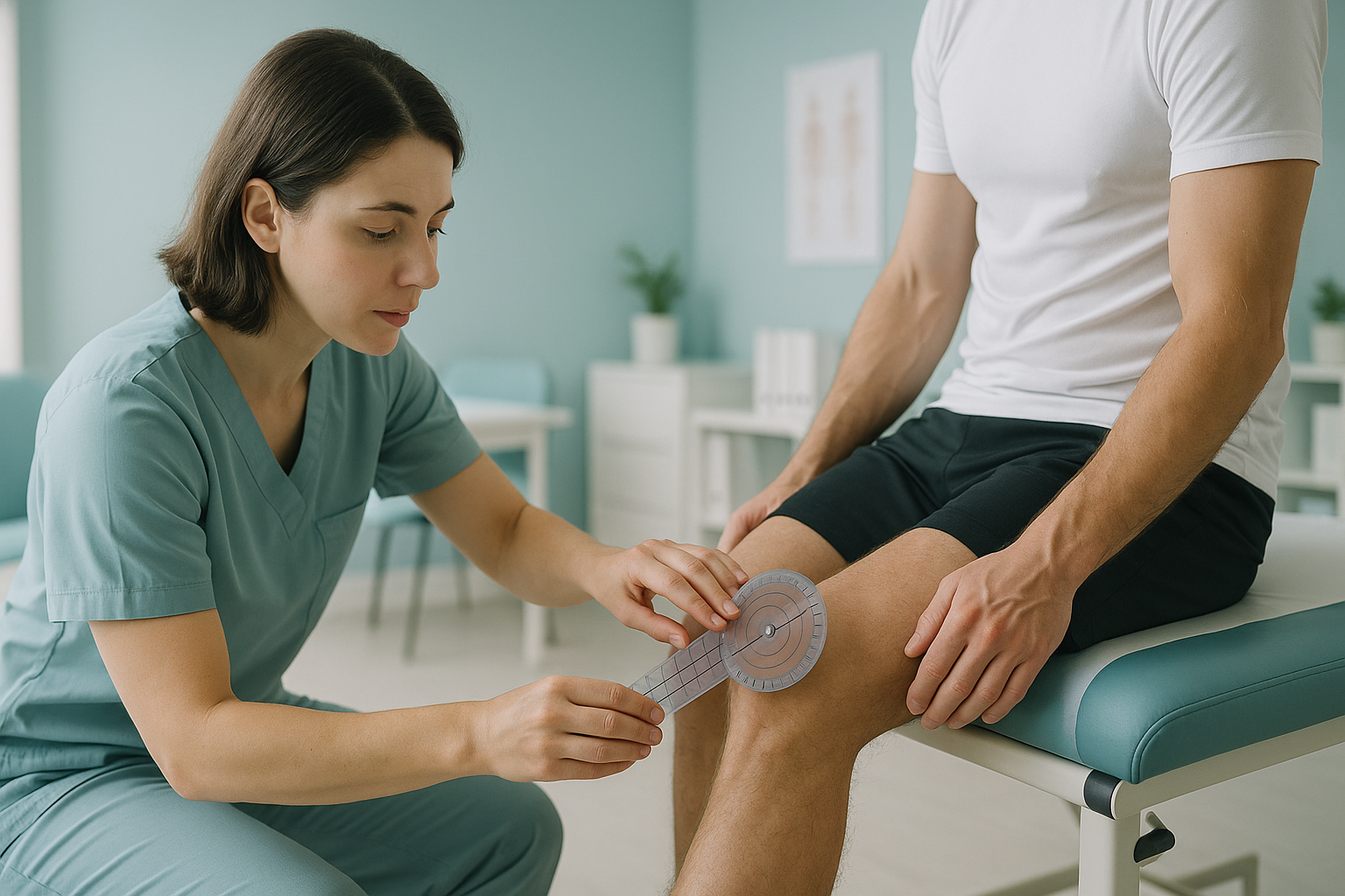
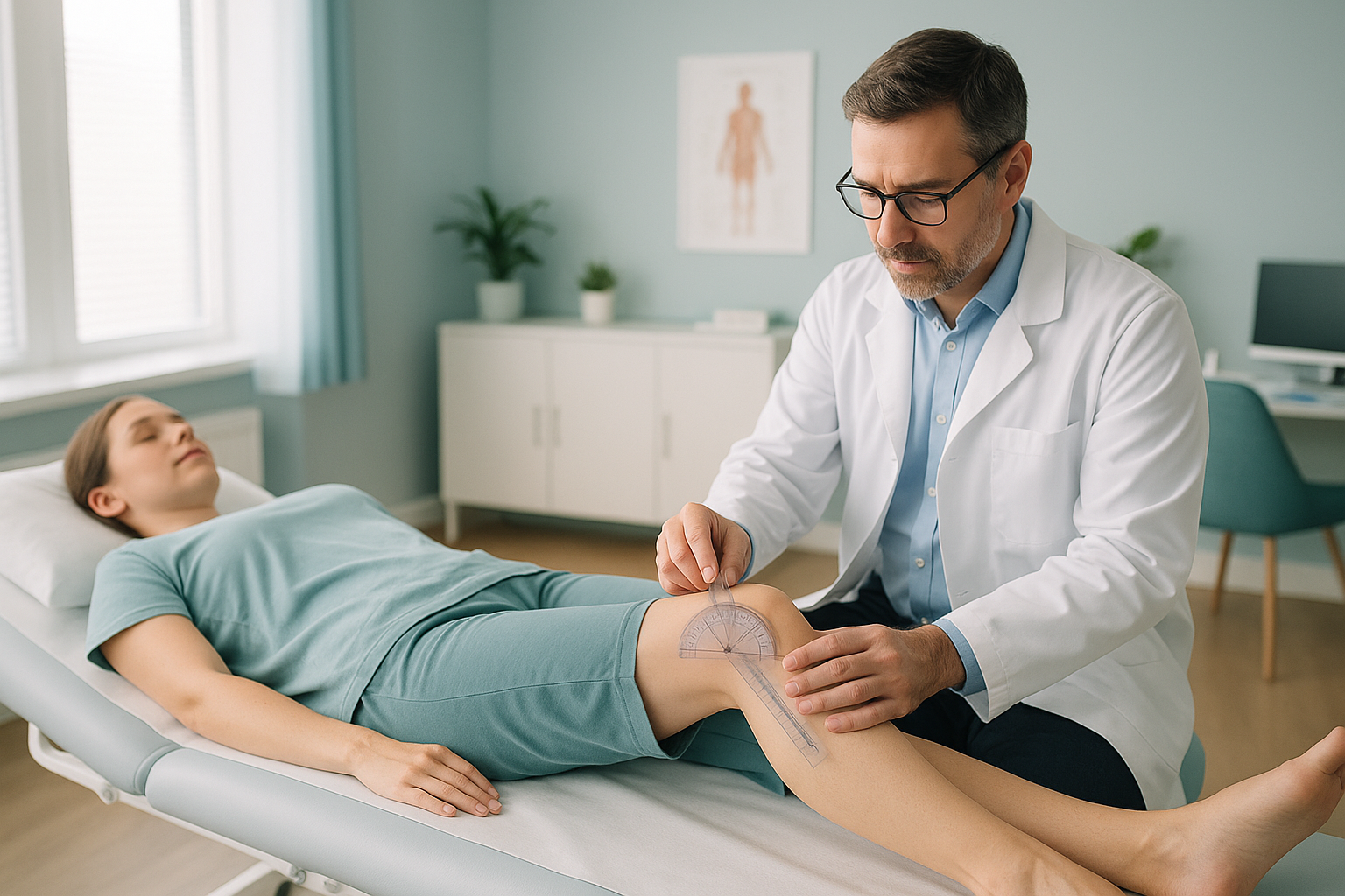
The Q angle is the angle between two imaginary lines. The first runs from the bony point at the front of your hip (the ASIS) down to the centre of your kneecap. The second runs from the centre of your kneecap down to the bump at the top of your shin (the tibial tuberosity). Where these two lines meet at the kneecap, they form the quadriceps angle.
Clinicians have traditionally measured it with a protractor while the patient lies flat or stands still. The idea is simple. A larger angle means the quadriceps pull the kneecap outward with more force. This could lead to poor patellar tracking and pain at the front of the knee.
Textbooks often quote a "normal" range that is somewhat higher in women than in men, mostly because women have a wider pelvis. For years, a higher Q angle was used to explain why women get more pain at the front of the knee. That idea is now being seriously questioned.
Why is the Q angle considered unreliable for diagnosing knee pain?
The Q angle is considered unreliable for several well-documented reasons. It is a fixed measurement taken in one position, yet knee pain usually happens during movement. Research has found that the same person can give clearly different Q angle readings. It depends on whether they are lying down or standing, and on which clinician measures them.
Perhaps the biggest problem is that the Q angle ignores everything above and below the knee. Your hip rotation, femoral version (whether the thighbone twists inward or outward more than usual), foot posture and trunk alignment all affect how force passes through the kneecap. A single number at the knee cannot capture all of that. To understand how the twist of the thighbone affects the load on the knee, read about anteversion versus retroversion.
What actually causes pain at the front of the knee, if not the Q angle?
Pain at the front of the knee is usually the result of several factors working together, not a single measurement. The body works as a connected chain from feet to hips. When alignment is off at one level, another area makes up for it, and the kneecap often pays the price.
These factors often combine to produce patellofemoral pain syndrome, one of the most common causes of pain at the front of the knee. Focusing only on the Q angle risks missing the real cause.
How does standing imaging show what the Q angle misses?
Standing, weight-bearing imaging shows the alignment your body actually works with. When you lie down for a usual X-ray or MRI, gravity is taken away, muscles relax, and joints settle into positions they never hold in daily life. A tilted pelvis may flatten out. A twist through the leg may disappear. The result is an image that looks more normal than reality.
What it captures: Full leg alignment from hips to ankles under your real body weight. Pelvic tilt, rotation through the femur, knee alignment and foot position are all visible in one image.
An EOS scan uses about 90% less radiation than a standard X-ray, around ten times less. ScanAlign standing EOS imaging captures the whole picture in seconds while you stand naturally.
What it misses: When you lie flat, gravity no longer loads the joints. A difference in leg length, a slight pelvic tilt or a rotational alignment problem may all straighten out and look normal on the image.
This is the main weakness of measuring the Q angle while lying down. The number you get may not reflect what happens when you stand and walk.
When MRI is the better tool: EOS shows bones, joints and alignment. For soft-tissue problems such as cartilage damage behind the kneecap, ligament tears or nerve irritation, MRI is usually the better choice. Your clinician can advise which scan you need.
Who should consider a standing alignment scan instead of relying on the Q angle?
A full standing assessment is worth considering if your pain at the front of the knee has not improved after physiotherapy, or if your scans so far have only looked at the knee on its own. The aim is to see whether the knee is making up for a problem somewhere else in the chain.
- Ongoing pain at the front of the knee or around the kneecap
- Earlier scans showed "nothing wrong" but the pain continues
- Told your Q angle is high but not offered further tests
- Knee pain that gets worse with stairs, squatting or running
- Sudden knee locking or giving way (see your doctor promptly)
- Suspected ligament or meniscus tear (MRI is usually needed first)
- Recent injury with significant swelling
Before you start a treatment, or change the one you are on, it is worth getting a detailed diagnosis. Knowing whether the cause sits in the pelvis, the legs or the spine changes what will actually help.
Not sure whether a standing alignment scan is right for your knee? Talk it through with us free of charge before you plan a trip to London.
Book a free video consultationFrequently asked questions about the Q angle and knee pain
Textbooks usually describe a range that is somewhat higher in women than in men, because women have a wider pelvis. However, there is a lot of natural variation, and the number on its own does not reliably predict whether someone will have knee pain.
Not necessarily. Large studies have found no consistent link between a higher Q angle and knee pain. Other factors, including hip strength, femoral version and movement patterns, play a much bigger role.
Women generally have a wider pelvis compared with their height, which increases the angle between the hip and the kneecap. This difference in body shape is normal and does not automatically mean a greater risk of knee problems.
The Q angle is mostly set by bone structure, so exercise will not change the angle itself. However, strengthening the hips and improving movement patterns can significantly reduce the abnormal forces that cause kneecap pain. This is usually more important than the angle.
Some clinicians still measure it as one of many data points, but it is rarely used as a diagnostic tool on its own. Modern practice puts far more weight on hip strength testing, movement analysis and imaging of the alignment of the whole leg.
Patellar maltracking means the kneecap does not glide smoothly in its groove during movement. A high Q angle was once thought to be the main cause. We now know maltracking can happen whatever the Q angle, often driven by hip control and soft-tissue balance. Read more about patellar maltracking.
An EOS scan captures the full skeleton from feet to hips (or higher) while you stand. This shows true weight-bearing alignment, including leg rotation, pelvic tilt and joint spacing, all in one image with about 90% less radiation than a standard X-ray, around ten times less.
No. ScanAlign is a self-referral clinic. You can book a measured alignment scan directly without a letter from your doctor.
Yes. EOS uses about 90% less radiation than a standard X-ray, around ten times less. It is one of the lowest-dose skeletal imaging systems available.
ScanAlign is a private, self-pay clinic. The price for patients travelling from abroad is confirmed during the free video consultation, where you can also discuss whether a scan is right for you. The consultation is free and with no obligation.
See the full picture, not just one angle
A free video consultation lets you discuss your knee pain with a specialist radiographer from home, and find out whether a standing alignment scan in London could help.
Book a free video consultationSources
- Smith TO, et al. "The Q angle: a systematic review." The Knee. 2011;18(3):166-171. PubMed.
- Powers CM. "The influence of abnormal hip mechanics on knee injury: a biomechanical perspective." Journal of Orthopaedic & Sports Physical Therapy. 2010;40(2):42-51.
- NHS. "Knee pain." nhs.uk. Accessed 2024.
Written by Abbas Dhami (Specialist Diagnostic Radiographer)
This page is general information and not a diagnosis.
ScanAlign is the trading name of CoreMed Solutions Ltd and operates under The Harley Street Hospital's CQC licence.