
Why Does Dose Matter Most in Paediatric Imaging?
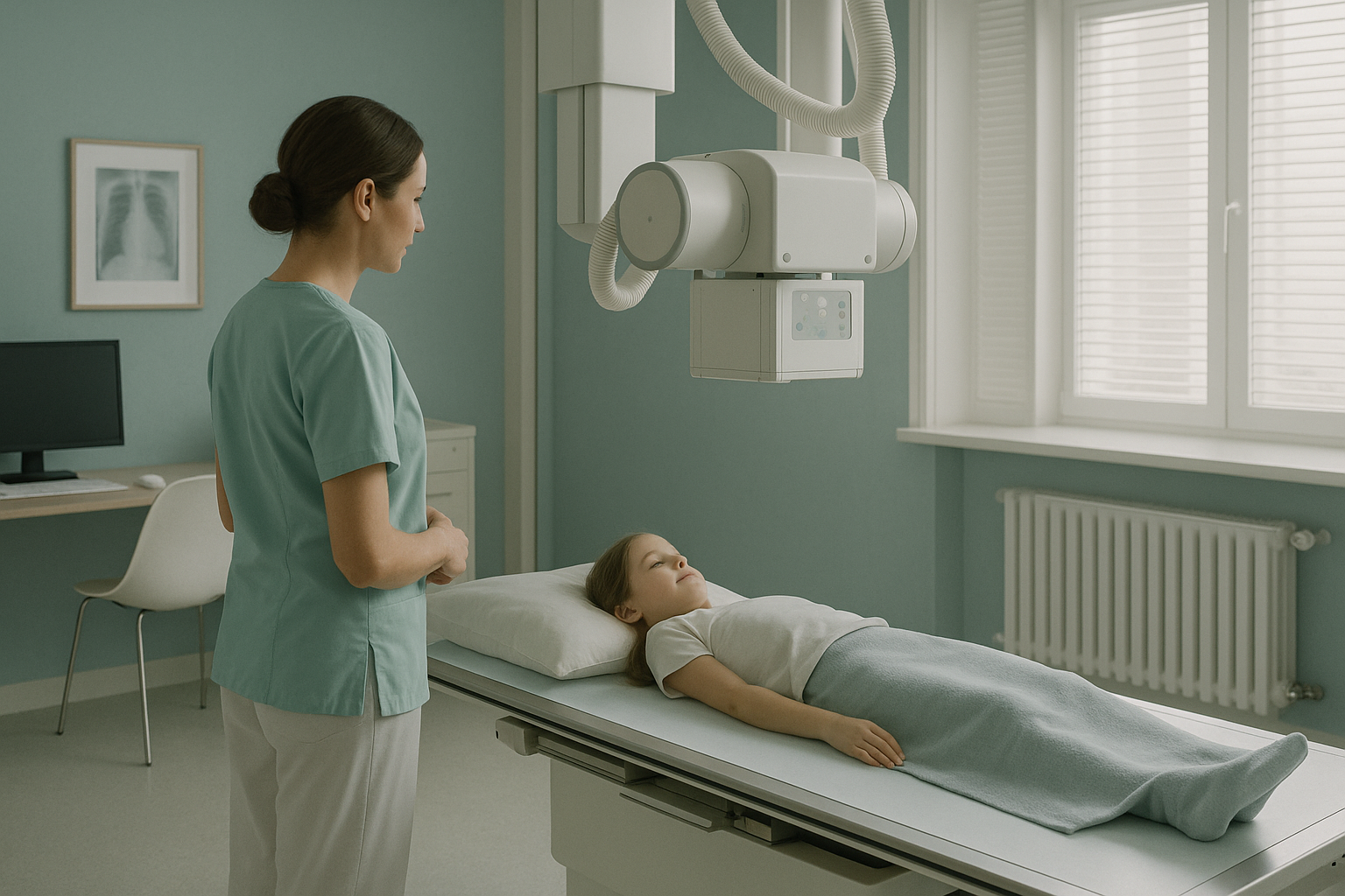
Children's cells divide quickly, so they are much more radiosensitive than adult tissue. The ICRP estimates that the radiation risk for a child under ten may be two to three times higher than for an adult given the same dose. For conditions that need repeated imaging, such as scoliosis monitoring, the total exposure becomes a real clinical concern. Low-dose methods like EOS can cut the dose by about 90% compared with standard radiography.
Key Takeaways
- Because children's tissue is more radiosensitive, keeping the dose low is a clinical priority, not just good practice.
- EOS gives about 90% less radiation than a standard X-ray, around ten times less, while capturing full standing alignment.
- Standing, weight-bearing imaging removes the need for sedation and gives a true functional picture of alignment.
- A parent or carer can stay in the room for the whole scan.
- ScanAlign accepts direct referrals from paediatricians, orthopaedic surgeons and general practitioners.
Why is radiation dose more important in children than in adults?
Children are not small adults. Their cells divide faster and their organs are still developing. They also have more years ahead in which an effect caused by radiation could, in theory, show up. The International Commission on Radiological Protection (ICRP Publication 103, 2007) estimates that the lifetime attributable risk of cancer per unit dose for a young child may be two to three times that of a middle-aged adult.
This does not mean a single X-ray is dangerous. It means that when imaging is clinically justified, the ALARA principle (As Low As Reasonably Achievable) should guide the choice of method. This matters most for conditions that need repeated exposure over months or years.
How does cumulative radiation exposure affect children with scoliosis?
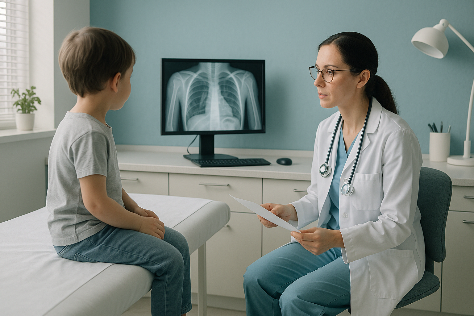
The total exposure over time is the real concern. A child diagnosed with scoliosis at age ten may have imaging every six to twelve months through adolescence. With standard radiography, that could mean ten or more full-spine exposures before the skeleton is mature.
Doody et al. (2000), in a large cohort study published in Spine, found a small but statistically significant rise in breast cancer deaths among women who had been imaged many times for scoliosis during childhood and adolescence using conventional radiography. The study looked at over 5,500 women and is still one of the most cited papers on this topic.
Deschênes et al. (2010), writing in the European Spine Journal, showed that EOS imaging lowered the effective dose for paediatric scoliosis studies by a factor of about six to nine compared with computed radiography, depending on patient size and protocol.
The clinical message is simple. When repeated imaging is needed, choosing the lowest-dose method that still answers the clinical question is more than following ALARA. It is measurably better care.
Scoliosis, limb length and follow-up after surgery often need imaging every 6 to 12 months.
Rapidly dividing cells in children are two to three times more radiosensitive than adult tissue (ICRP 103).
Low-dose EOS gives about 90% less radiation than a standard X-ray, around ten times less.
For more on dose reduction and the technology behind it, see EOS scan safety and radiation.
Why does standing imaging give a better clinical picture in children?
Standing, weight-bearing imaging shows the skeleton under the same gravitational load the child carries every day. This matters because the body is a connected chain from feet to head. A problem in one area often makes another area compensate. Lying-down imaging can relax the muscles and hide alignment problems that only show under upright load.
For scoliosis, a supine MRI or lying-down X-ray may underestimate how severe the curve is, because gravity and muscle tone are taken out of the picture. An EOS scan captures the full spine and lower limbs in one standing exposure, giving you a true functional picture of alignment.
Weight-bearing alignment: Gravity loads the skeleton naturally. This shows the true size of the curve, pelvic obliquity (uneven pelvis) and limb length differences as the child actually lives with them.
No sedation needed: The child simply stands still for a few seconds. A parent or carer stays in the room the whole time.
Full kinetic chain in one image: Head to feet in a single low-dose exposure, so you see the whole compensation pattern at once.
Muscles relaxed: Lying down removes the load of gravity. Curves, tilts and compensations may look less severe than they are in daily function.
Sedation sometimes needed: Young children who cannot stay still for an MRI or CT may need sedation. This adds risk and makes things more complicated to arrange.
Imaging in sections: Conventional X-rays often image the spine in parts. This makes a full alignment assessment harder and needs the images to be stitched together.
You can read more about paediatric imaging at ScanAlign, including the child-friendly setting and what families can expect on the day.
How do I refer a child for an EOS scan?
ScanAlign accepts direct referrals from paediatricians, orthopaedic surgeons, physiotherapists and general practitioners. No go-between is needed. The process is designed to be quick and simple.
If you have not referred before, register to refer patients. It takes a few minutes and you only need to do it once.
Fill in the online referral form with the clinical question and relevant history. State the region you need imaged (full spine, lower limbs, or both).
The family comes to 19 Harley Street, London. A parent or carer stays with the child the whole time. The scan takes seconds and no sedation is needed.
A detailed report with full alignment analysis is sent directly to you, the referring clinician. The images are available on a secure platform.
Ready to refer a child? You can also see what ScanAlign, low dose standing imaging offers for all age groups.
When should I choose EOS over MRI for a child?
EOS and MRI answer different clinical questions. The right choice depends on what you need to see.
- Scoliosis monitoring and curve progression
- Limb length discrepancy assessment
- Lower limb alignment (valgus, varus, torsion)
- Review of implants after surgery
- Pelvic obliquity and overall balance
- Repeated follow-up where total dose is a concern
- Soft tissue, disc or nerve assessment
- Suspected spinal cord abnormalities
- Bone marrow disease or infection
- Soft tissue injuries inside the joint
- Conditions that need contrast
A note on MRI: EOS shows bones, joints and alignment under weight-bearing load. For soft tissue, discs or nerves, MRI is usually the better tool. The two work together, and in many paediatric musculoskeletal cases both may be right at different stages of assessment.
Before a child starts treatment, or changes the one they are on, it is worth getting a detailed diagnosis. Knowing whether the cause sits in the pelvis, the legs or the spine changes what will actually help.
Frequently asked questions from referring clinicians
Yes. EOS gives about 90% less radiation than a standard X-ray, around ten times less. Published data from Deschênes et al. (2010) confirms dose reductions of six- to nine-fold for paediatric spine studies compared with computed radiography. For more detail, see our page on EOS scan safety and radiation.
Yes. If you have not referred before, you can register to refer patients online. It takes a few minutes and you only need to do it once.
Yes. A parent or carer stays in the room with the child for the whole scan. The child simply stands still for a few seconds. No sedation is needed.
ScanAlign can image children who are able to stand still for a few seconds. The minimum age depends on how well the child can cooperate. There is no strict lower limit, but most referrals are for school-age children and adolescents. We also scan adults.
Reports include a full standing alignment analysis with calibrated measurements, annotated images, and a written commentary on the clinical question you gave in the referral. Images are available on a secure platform.
ScanAlign is a private, self-pay clinic. The price for patients travelling from abroad is confirmed during the free video consultation.
Yes. Scoliosis monitoring is one of the strongest clinical reasons to use EOS. Because the dose is low, repeated imaging over months or years adds up to a much lower total exposure than conventional radiography. Standing images also give a functional, weight-bearing assessment of how severe the curve is.
EOS and MRI work together. One cannot replace the other. EOS excels at bony alignment under load. MRI is better for soft tissue, discs, nerves and bone marrow disease. In many cases, both may be right at different stages.
Children are usually asked to change into light clothing with no metal. The team will explain what to wear when the appointment is confirmed. The aim is to keep the child comfortable and at ease.
ScanAlign is at 19 Harley Street, London, inside The Harley Street Hospital. The clinic is easy to reach by public transport in London.
Refer a child
If you have a young patient who needs low-dose standing alignment imaging, we are here to help.
Book a free video consultationSources
- International Commission on Radiological Protection. The 2007 Recommendations of the International Commission on Radiological Protection. ICRP Publication 103. Ann ICRP. 2007;37(2-4):1-332.
- Deschênes S, Charron G, Beaudoin G, et al. Diagnostic imaging of spinal deformities: reducing patients radiation dose with a new slot-scanning X-ray imager. Eur Spine J. 2010;19(1):118-126.
- Doody MM, Lonstein JE, Stovall M, Hacker DG, Luckyanov N, Land CE. Breast cancer mortality after diagnostic radiography: findings from the U.S. Scoliosis Cohort Study. Spine. 2000;25(16):2052-2063.
Written by Abbas Dhami (Specialist Diagnostic Radiographer)
This page is general information and not a diagnosis.
ScanAlign is the trading name of CoreMed Solutions Ltd and operates under The Harley Street Hospital's CQC licence.