
How Does Rugby Load Affect the Spine Over a Career?
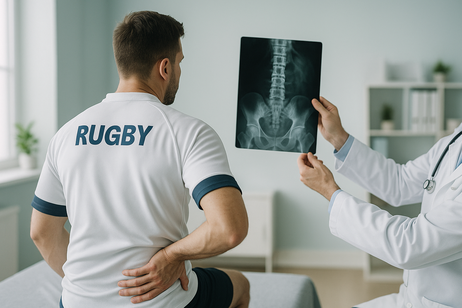
Rugby puts a huge amount of load on the spine over time, through scrums, tackles and collisions. Over a playing career, this repeated stress can lead to early wear in the discs and joints of the neck and back. Many of these changes only show up when the spine is checked while you stand, carrying your own body weight.
Key points
- Scrums and tackles put the cervical and lumbar spine under repeated high forces.
- Front row forwards face especially high neck loads, but every position is affected.
- Load that builds up over time can cause early degenerative changes in discs and joints.
- Scans taken lying down can miss alignment problems that only show under gravity and body weight.
- A standing full spine X-ray shows how the whole chain is coping with years of load.
A sudden neck or spine injury in rugby is a medical emergency. If you or someone else has sudden neck pain after a tackle, scrum collapse or collision, especially with numbness, tingling, weakness in the arms or legs, or any loss of bladder or bowel control, do not move. Call your local emergency number or go to the nearest emergency department straight away. This page is about long-term spinal changes that build up over time, not sudden injuries.
What happens to a rugby player's spine over time?
Rugby is a collision sport. Every scrum, every tackle and every ruck adds load to the vertebrae, discs and ligaments. On its own, each of these forces is usually well within what a healthy spine can handle. But the total load over seasons and decades slowly speeds up natural wear and tear.
Research on retired professional players shows higher rates of early disc narrowing, joint stiffness and bony changes than in non-contact athletes of the same age. These are the same processes seen in degenerative disc disease and cervical spondylosis. In rugby players, though, they tend to appear earlier and affect more levels of the spine.
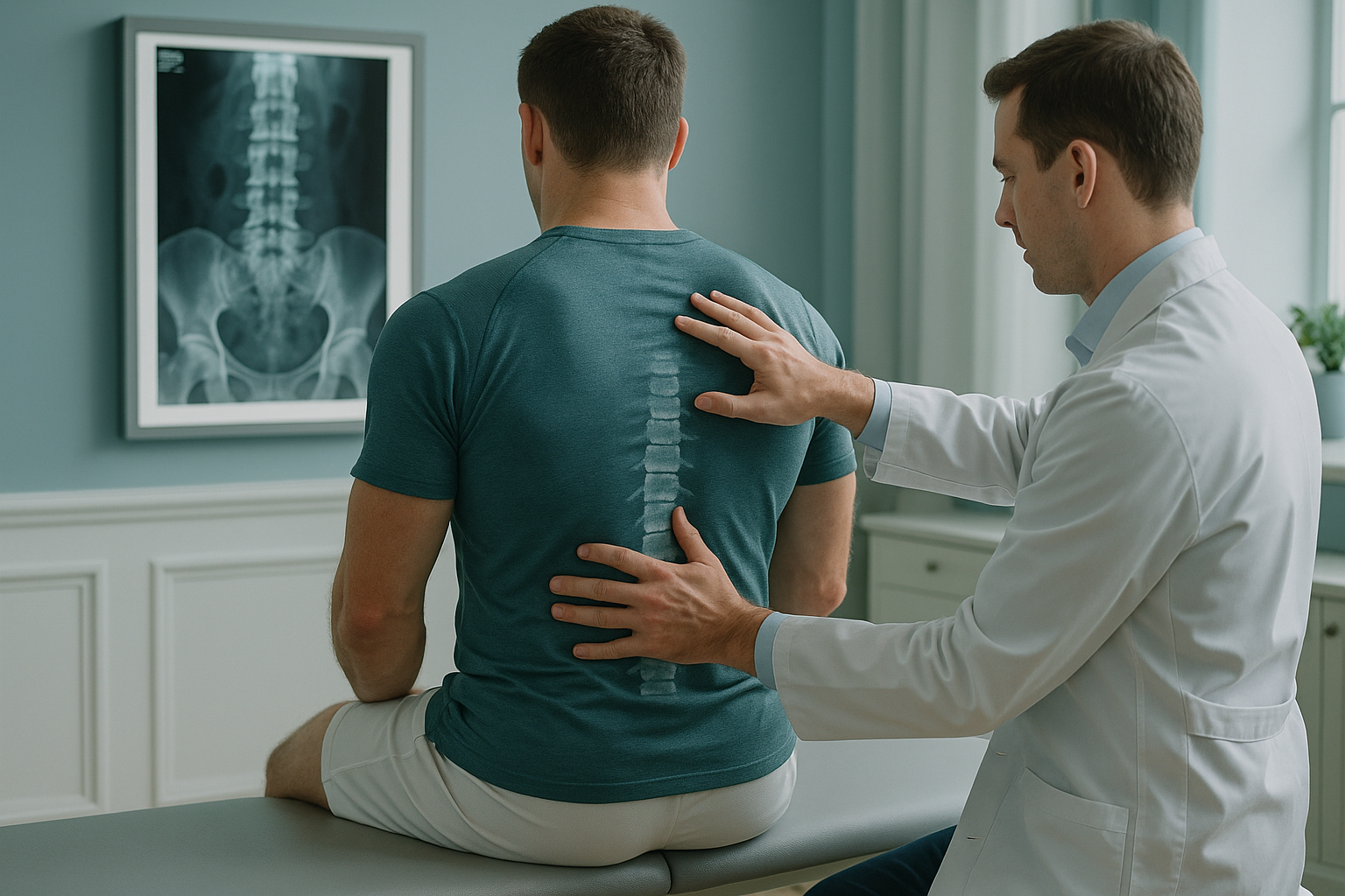
The neck is especially at risk. During a scrum, the cervical spine takes compressive and shear forces that can be several times body weight. Over thousands of scrums in a career, the discs and facet joints in the neck change shape. This process is not always painful at first. That is why many players only notice stiffness and less movement years after they retire.
What are the signs of long-term rugby spine injury?
Spinal changes that build up over time can develop slowly. You may notice some of these during your career or after you retire.
What causes degenerative changes in the spine from rugby?
No single event is to blame. What matters is the total load across a career. Several factors work together.
Your body works as a connected chain. When the cervical spine stiffens, the thoracic and lumbar regions make up for it. When the lower back takes more load than it should, the hips and pelvis adjust. Pain often ends up appearing away from the area where it all started. That is why scans of just one region can miss the bigger picture.
Why does a standing scan show what lying-down imaging misses?
Most scans ask you to lie flat. When you lie down, gravity is taken out of the picture. Your muscles relax, your discs decompress slightly, and mild misalignments can disappear from the image. The result shows your spine at rest, not your spine under real, everyday load.
A standing, weight-bearing EOS scan captures your whole spine and pelvis in one image while you stand naturally. It shows how gravity, muscle tone and years of rugby load have shaped your alignment from head to pelvis. ScanAlign standing EOS imaging uses about 90% less radiation than a standard X-ray, around ten times less. That makes it practical for monitoring over time, not just a single snapshot.
Wondering how your spine has handled years of rugby? A free video consultation can help you decide whether a standing scan in London is the right next step, before you plan any travel.
Book a free video consultationHow can rugby players keep track of their spine health?
Good sports injury prevention is not only about avoiding the big injury. It is also about tracking the slow changes that build up. A baseline standing scan gives you and your clinician a clear picture of where your spine is now. If you have another scan in future, changes can be measured against that baseline.
This kind of monitoring is especially useful for front row forwards with neck stiffness, retired players whose back pain is getting worse, or anyone whose symptoms do not match what a lying-down MRI shows. It can also guide strength training by showing which regions are under the most strain.
If your clinician needs a closer look, you can book a standing spine scan at our clinic at 19 Harley Street, London. The scan itself takes only a few minutes.
Before you start treatment, or change the one you are on, it is worth getting a detailed diagnosis. Knowing whether the cause sits in the pelvis, the legs or the spine changes what will actually help.
When MRI is the better choice: EOS shows bones, joints and alignment. For soft tissue problems such as disc bulges pressing on nerves, ligament tears or checking the spinal cord, MRI is usually the better tool. Many rugby players benefit from both.
Frequently asked questions about rugby and spinal load
Rugby is a collision sport. Research shows that retired players have higher rates of early disc and joint wear than non-contact athletes. This does not mean every player will have problems, but the load that builds up over time does increase the risk of earlier degenerative changes.
Yes. Front row players take the highest compressive forces through the cervical spine during scrums. Studies consistently show higher rates of neck stiffness and cervical degeneration in props and hookers than in backs.
A sudden (acute) injury happens all at once, often from a scrum collapse or an awkward tackle. It can involve fractures, dislocations or spinal cord damage. That is a medical emergency. Long-term wear is a slow process where discs narrow and joints stiffen over years of repeated load. It may not cause symptoms until well into a career, or after it.
A standing EOS scan can show joint narrowing, bony spurs and alignment changes that are linked with early arthritis. Importantly, it shows these under real weight-bearing conditions. This can reveal problems that look less significant when you are lying flat.
An EOS scan uses about 90% less radiation than a standard X-ray, around ten times less. This low dose makes it practical for repeat monitoring scans over time.
No. You can refer yourself. The process starts with a free video consultation. A specialist radiographer talks through your history and symptoms and advises whether a scan is right for you.
The price for patients travelling from abroad is confirmed during the free video consultation.
Either can help. A scan during your career can set a baseline and guide your strength and conditioning training. A scan after retirement can make clear the source of any lingering stiffness or pain.
EOS can show narrowing of the disc space and overall alignment, which are strong signs of disc wear. For a detailed soft tissue check of the discs, including bulges and nerve compression, MRI is usually more suitable.
Absolutely. Amateur players who have trained and played regularly for years build up similar types of spinal load. If you have persistent back or neck stiffness from rugby at any level, a standing alignment assessment can be valuable.
See how your spine has handled years of rugby
The first step is a free video consultation with a specialist radiographer, from wherever you are. No referral needed, no obligation.
Book a free video consultationSources
- Castinel, B.H. et al. "Cervical spine injuries in French rugby union players." British Journal of Sports Medicine, 2009; 43(13): 1024-1028.
- Hogan, B.A. et al. "The cervical spine of professional front-row rugby players: correlation between degenerative changes and symptoms." Irish Journal of Medical Science, 2010; 179(2): 259-263.
- World Rugby. Scrum Engagement Law Amendments and Player Welfare. World Rugby Player Welfare resources.
Written by Abbas Dhami (Specialist Diagnostic Radiographer)
This page is general information and not a diagnosis.
ScanAlign is the trading name of CoreMed Solutions Ltd and operates under The Harley Street Hospital's CQC licence.