
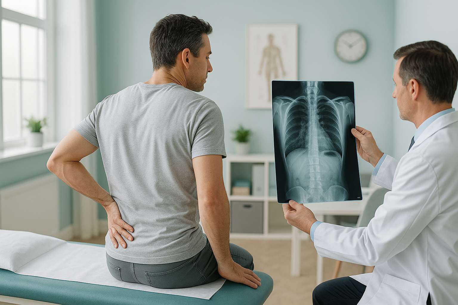
When Does Chronic Back Pain Actually Need a Scan?
Most back pain settles within a few weeks and does not need a scan. A scan becomes truly useful in three cases: when pain has lasted beyond six to twelve weeks despite conservative care, when specific red-flag symptoms are present, or when a specialist needs structural detail before recommending the next step in your care.
Key points
- Guidelines recommend trying conservative management first for most back pain.
- Red-flag symptoms such as numbness, weakness or bladder changes need urgent medical attention, not a routine scan booking.
- After six to twelve weeks of ongoing pain, a scan can help find a structural cause.
- Standing scans show your spine under real load, which lying-down scans can miss.
- A scan is a diagnostic tool, not a treatment. It guides the next decision.
If you have lived with back pain for months, you have probably asked yourself whether you need a scan. The honest answer is: it depends. Scanning too early can show findings that look alarming but are completely normal for your age. Scanning at the right time, with the right method, can change everything about how your problem is understood and managed.
This page takes you through the guidelines, the red flags and the situations where a scan really moves you forward. We also explain why the position you are scanned in matters more than most people realise.
Why do guidelines say not to scan back pain straight away?
Because most back pain gets better on its own, and early scans rarely change the outcome. In the UK, both NICE and the Royal College of Radiologists advise against routine scans for uncomplicated lower back pain in the first six weeks. The reasons are simple.
Around 90% of acute back pain episodes clear up within 6 to 12 weeks with movement, gentle exercise and pain management.
Scans often show disc bulges or wear that look worrying but are normal age-related changes and may have nothing to do with your pain.
If a scan is done too soon, there is a risk of treating the image rather than the person. This can lead to procedures you may not need.
This does not mean your pain is not real. It means that in the early weeks, conservative management, staying active and working with a physiotherapist, is the recommended first step.
What are the red-flag symptoms that need urgent attention?
Some symptoms mean you should get medical help immediately, not book a routine scan. If you have any of the following, contact your doctor or go to your nearest emergency department.
This can be a sign of cauda equina syndrome, a medical emergency that needs assessment the same day.
Nerve symptoms that spread or get worse over days need a prompt clinical examination.
These may signal infection or another serious condition that needs investigation beyond a scan.
Numbness around the groin or inner thighs is another sign of possible cauda equina syndrome. Get help the same day.
If any of these apply to you, please stop reading and contact a medical professional now. The rest of this page is for people with chronic, persistent back pain without these urgent signs.
When does a scan for chronic back pain actually help?
A scan becomes useful when your pain has lasted beyond the expected recovery time and you need clearer answers. These are the most common situations where a scan adds real value.
- Pain lasting beyond 6 to 12 weeks despite physiotherapy, exercise and proper pain management.
- Your specialist or surgeon needs structural detail before recommending an injection, a procedure or a referral. Read more about when to see a specialist.
- A previous scan left questions unanswered, especially if it was taken lying down and your pain is mainly when you stand or walk.
- You are considering surgery and your surgeon needs weight-bearing images to plan the approach.
- You want a second opinion on a diagnosis and need fresh, detailed images to take to another clinician.
In each of these cases, the scan is not the treatment. It is the tool that helps you and your clinician decide what to do next.
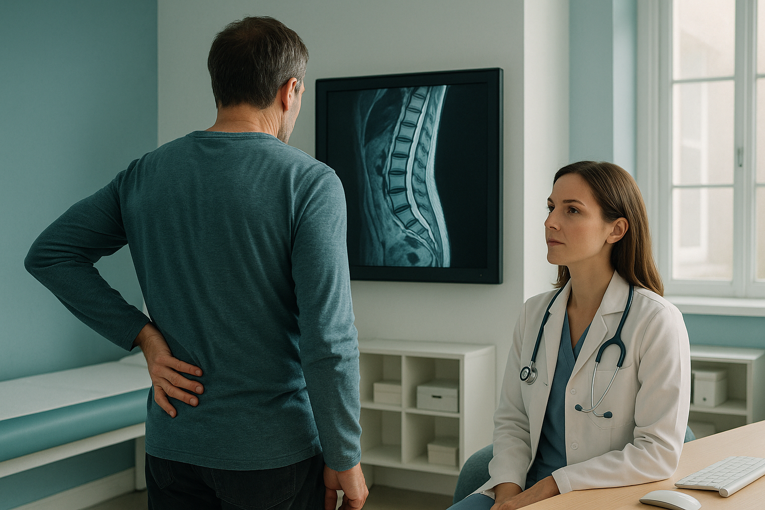
Not sure if a scan is the right step for you? You can check whether a scan is right for you with a free video consultation from home. There is no pressure to book.
Book a free video consultationWhy does the position you are scanned in matter?
It matters because your spine behaves differently when you stand under gravity than when you lie on a scanner bed. Most conventional scans, including standard X-rays, CT and MRI, are done with you lying flat. In that relaxed position your muscles switch off, your curves settle and your joints decompress.
Standing scans capture your skeleton while it carries your full body weight. This shows how your vertebrae actually take load, whether your pelvis tilts, and how your curves shift to compensate. Problems like spondylolisthesis (where one vertebra slips forward), subtle scoliosis or load-related narrowing often become visible only in this position.
Lying-down scans are excellent for many things, but they take gravity out of the picture. A slipped vertebra can move back to a normal position. A curve can flatten out. Your report may come back "normal" even though you feel anything but normal when you are on your feet.
Your body works as a connected chain from your feet to your head. A tilt in the pelvis, a difference in leg length or a stiff hip can each force your spine to compensate. Pain often appears away from the root cause. A whole-body standing image, like the one EOS produces, captures that full chain in a single exposure with about 90% less radiation than a standard X-ray, around ten times less.
Learn more about ScanAlign, private standing imaging at 19 Harley Street, London, and find out what to expect at an alignment assessment.
Should I get an X-ray or MRI for back pain?
It depends on what your clinician is looking for. Each type of scan answers a different question.
- Standing X-ray or EOS scan
- Shows vertebral position, disc height, spinal curves, pelvic balance, leg alignment
- Best for structural and load-related problems
- MRI scan
- Shows disc herniation, nerve compression, inflammation, soft tissue damage
- Best for suspected nerve involvement or disc problems
A note on MRI. EOS and standing X-rays show bones, joints and alignment very clearly. For soft tissue, discs or nerves, MRI is usually the better tool. Sometimes you need both. A free video consultation can help you understand which type of scan, if any, is the right starting point for you.
For more detail on what our clinic covers, visit our back pain clinic page.
What happens if I decide to get a scan?
If you choose to look into a scan with us, the process is simple and you do not need a referral.
A short call to understand your history, your symptoms and whether a scan is likely to help. If it is not the right step, we will tell you honestly.
A quick, comfortable scan at 19 Harley Street, London. You stand naturally while the system captures a full-body image from head to feet.
Your images are analysed and a detailed report is prepared. It covers alignment, balance and any structural findings relevant to your symptoms.
You own your images and report. Share them with your doctor, physiotherapist, osteopath or surgeon back home to guide the next stage of your care.
Before you start a treatment, or change the one you are on, it is worth getting a detailed diagnosis first. Knowing whether the cause sits in your pelvis, your legs or your spine changes what will actually help.
Frequently asked questions
No. ScanAlign is a private self-pay clinic. You can book directly without a referral from your doctor. However, we always recommend keeping your doctor informed about any scans you have.
Guidelines generally suggest trying conservative management for six to twelve weeks first. If pain continues after that, or if your clinician suspects a specific structural problem, a scan becomes more useful.
It depends on the question being asked. X-ray and EOS are best for bone position, alignment and structural balance. MRI is best for soft tissue, discs and nerves. Sometimes both are needed.
An EOS scan captures your entire skeleton while you stand, from head to feet, in a single exposure. It uses about 90% less radiation than a standard X-ray, around ten times less. Because you are upright, it shows how your bones and joints behave under real body weight.
A scan shows structural detail, such as how your spine curves, whether your pelvis is level, or if a vertebra has shifted. It does not diagnose the pain itself, but it gives your clinician the information they need to make a more informed decision about your care.
When you lie down, gravity is removed and your muscles relax. A vertebral slip can reduce, a curve can flatten, and joint spacing can change. Problems that only appear under load may not show up at all when you are lying down.
The price for patients travelling from abroad is confirmed during your free video consultation.
Yes. The EOS system uses about 90% less radiation than a standard X-ray, around ten times less. It is one of the lowest-dose imaging methods available for full-body skeletal assessment.
If you have progressive numbness, weakness in your legs, or changes to bladder or bowel control, please get urgent medical attention. Contact your doctor or go to your nearest emergency department. These symptoms need assessment the same day, not a routine scan booking.
It is a short, no-pressure call. We ask about your symptoms, your history and what you hope to learn. If a scan is not the right step for you, we will say so. If it is, we explain exactly what the process involves and what the images can show.
Not sure if a scan is right for you?
Book a free, no-pressure video consultation before you plan any travel. We will be honest about whether a scan will actually help in your situation.
Book a free video consultationWritten by Abbas Dhami (Specialist Diagnostic Radiographer)
Sources
- NICE Guideline NG59: Low back pain and sciatica in over 16s: assessment and management. National Institute for Health and Care Excellence, 2016 (updated 2020).
- The Royal College of Radiologists. iRefer: Making the best use of clinical radiology. Guidelines on appropriate imaging referrals.
- Brinjikji W, Luetmer PH, Comstock B, et al. Systematic literature review of imaging features of spinal degeneration in asymptomatic populations. AJNR Am J Neuroradiol. 2015;36(4):811-816.
This page is general information and not a diagnosis.
ScanAlign is the trading name of CoreMed Solutions Ltd and operates under The Harley Street Hospital's CQC licence.