
Scanogram or EOS: Which Full-Leg Measurement Is More Accurate?
A scanogram is a long-leg X-ray. It is used to measure leg length and alignment. EOS takes the same measurements in one pass while you stand. It uses about 90% less radiation than a standard X-ray, around ten times less. EOS images the whole leg in one sweep. This means it avoids the stitching errors and magnification problems that can affect traditional scanograms.
Key takeaways
- A scanogram joins several X-ray shots together. If the patient moves, this can cause measurement errors.
- EOS uses slot-scanning technology. It images both legs in one continuous pass while you stand.
- Standing imaging shows how your bones and joints behave under your own body weight. Scans done lying down cannot show this.
- EOS uses about 90% less radiation than a standard X-ray, around ten times less.
- Both methods have a place. But EOS reduces the main causes of inaccuracy in full-leg X-rays.
What is a scanogram and how does it work?
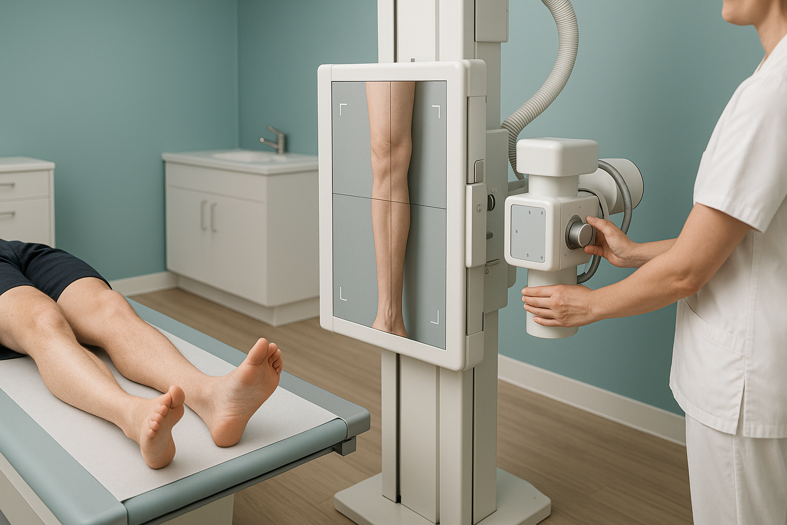
A scanogram is a type of full-leg standing X-ray. It measures the length and alignment of your legs. The traditional method takes three separate X-ray shots: one at the hip, one at the knee and one at the ankle. These are then joined digitally into one long image.
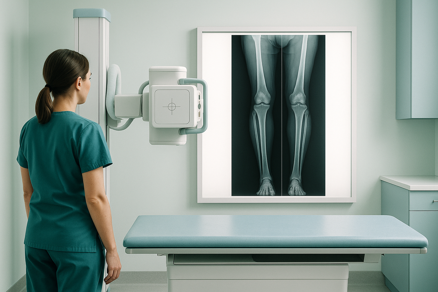
This joined image is sometimes called a long leg film. On it, a clinician can draw measurement lines from the hip joint to the ankle. This checks for differences in leg length or in the angle of alignment. Scanograms have been used reliably for decades. Orthopaedic surgeons still often ask for them.
However, the image is built from separate shots, each taken at a slightly different X-ray beam angle. Because of this, two well-known problems can creep in: magnification distortion and stitching artefact.
Why are scanogram measurements sometimes less accurate?
Several technical factors can affect scanogram measurements. None of them make a scanogram useless. But they explain why surgeons more and more ask for EOS when precision matters most.
If you shift even slightly between shots, the images may not line up perfectly where they join. This mismatch can add to or take away from the true length.
Standard X-rays use a cone-shaped beam. Objects closer to the source look larger. Without careful calibration, bone lengths can be measured as too long or too short.
A full-leg scanogram can take several seconds across three shots. Any shift in weight or any turning during that time affects the final joined image.
EOS deals with these problems by using slot-scanning technology. A narrow fan-shaped beam sweeps from head to toe in one continuous pass. There is no stitching, because the image is taken in one go. The parallel beam also keeps magnification the same from top to bottom. So there is no need for calibration afterwards.
EOS scans you while you stand, so the image shows how your legs really carry weight. Lying down relaxes the muscles and can hide the true alignment. So a standing scan often tells a different story, one that is more useful to your clinician. Learn more about leg alignment and why a weight-bearing position matters.
How does EOS compare with a scanogram for full-leg imaging?
EOS and scanograms both produce a long image of the legs. But they differ in how that image is taken and what it can show. The table below sums up the key differences.
| Feature | Traditional scanogram | EOS |
|---|---|---|
| How the image is taken | Several shots joined together | One continuous slot scan |
| Patient position | Standing or lying (depends on the department) | Standing, weight-bearing |
| Risk of stitching artefact | Present at each join | None, one-pass image |
| Magnification | Cone-beam distortion, needs calibration | Minimal, close to 1:1 scale |
| Radiation | Standard X-ray dose | About 90% less than a standard X-ray |
| Front and side images at the same time (biplanar) | Not available | Front and side views taken at once |
| 3D reconstruction | Not possible from a single scanogram | Available from the biplanar data |
For a wider look at how EOS fits alongside other imaging methods, read our guide to EOS compared with X-ray, CT and MRI.
When MRI is the better choice: EOS shows bones, joints and alignment clearly. For soft-tissue detail, such as discs, cartilage, ligaments or nerves, MRI is usually the better tool. Your clinician can advise which scan you need.
How do I book a standing full-leg scan with EOS?
You can book an EOS standing X-ray yourself at ScanAlign, standing EOS imaging in London. You do not need a referral from your doctor. Here is what to expect.
Free video consultation
A specialist radiographer goes through your history with you. They confirm whether an EOS scan is the right next step for you.
Your scan at 19 Harley Street, London
You stand in the EOS booth in normal clothes (no metal). The scan takes under a minute. It takes front and side views at the same time.
Detailed report
Your images and measurements are put together in a clear report. You can share it with your treating clinician.
Next steps
You and your clinician use the report to guide any further decisions. ScanAlign is a diagnostic clinic, so treatment happens with the specialist you choose.
Before you start treatment, or change the one you are on, it is worth getting a detailed diagnosis. Knowing whether the cause sits in the pelvis, the legs or the spine changes what will actually help.
Ready to get accurate full-leg measurements with less radiation?
Book a free video consultationFrequently asked questions
A scanogram is a full-leg X-ray. It is used to measure differences in leg length and overall leg alignment. Orthopaedic surgeons often ask for one before or after joint replacement or corrective surgery.
A CT scanogram is a scout image made by a CT scanner. It gives good length measurements. But it uses a higher radiation dose than a plain X-ray scanogram. It also images you lying down, so it does not show weight-bearing alignment.
EOS uses about 90% less radiation than a standard X-ray, around ten times less. A traditional scanogram gives a standard X-ray dose, so EOS is much lower.
When you stand, gravity and your muscles affect joint angles and alignment. Lying down relaxes everything. This can hide a tilt, a twist or a length difference that only shows up under load. Standing imaging captures what your body actually does when you are on your feet.
A traditional scanogram takes separate X-ray shots of the hip, knee and ankle, then joins them digitally. If you move between shots, the images may not line up perfectly. The mismatch at each join is called a stitching artefact. It can affect measurements.
No. ScanAlign accepts patients who book themselves. You start with a free video consultation. In it, a specialist radiographer checks whether the scan is suitable for you.
ScanAlign is a private, self-pay clinic. For patients travelling from abroad, the price is confirmed during the free video consultation, which you can arrange on the book a full-leg standing scan page.
Yes. EOS takes front and side views at the same time (simultaneous biplanar images). Because of this, the data can be used to build a 3D reconstruction of the shape of your bones and joints. A standard scanogram cannot do this.
EOS has clear advantages in radiation dose, stitching accuracy and weight-bearing position. A traditional scanogram is still a perfectly valid tool in many departments. Your surgeon may have good reasons for asking for one. The two are not competing in every situation.
You can wear normal clothes as long as they have no metal zips, buttons or fastenings in the scan area. The radiographer will advise you on the day and give you a gown if needed.
Get precise, low-dose full-leg measurements
A standing EOS scan captures your true alignment in one pass, with less radiation and no stitching artefact. Start with a free video consultation to see if it is right for you.
Book a free video consultationSources
- Deschênes S, Charron G, Beaudoin G, et al. "Diagnostic imaging of spinal deformities: reducing patients' radiation dose with a new slot-scanning X-ray imager." Spine, 2010;35(9):989-994.
- Illés T, Somoskeöy S. "The EOS™ imaging system and its uses in daily orthopaedic practice." International Orthopaedics, 2012;36(7):1405-1411.
- NHS England. "Diagnostic imaging dataset statistical release." Available at digital.nhs.uk.
Written by Abbas Dhami (Specialist Diagnostic Radiographer)
This page is general information and not a diagnosis.
ScanAlign is the trading name of CoreMed Solutions Ltd and operates under The Harley Street Hospital's CQC licence.