
When Is Scoliosis Surgery Recommended?
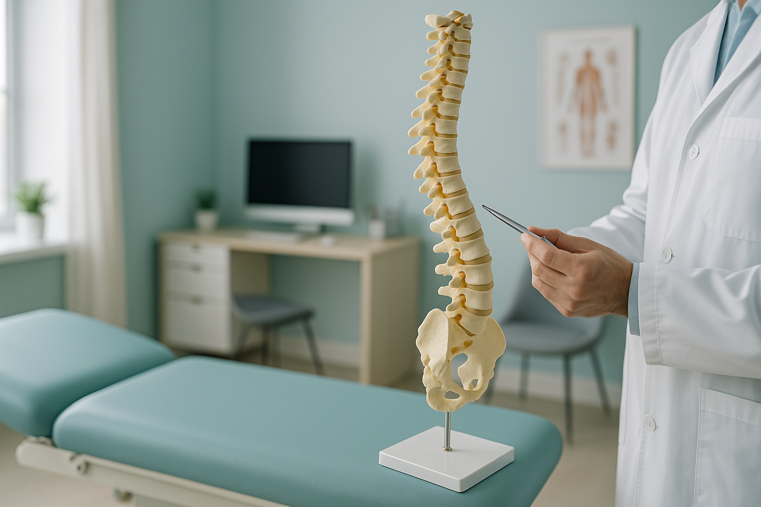
Scoliosis surgery is usually recommended when a spinal curve is large, progressing, or causing significant pain and reduced lung function that non-surgical treatments have not controlled. The decision depends on the size of the curve, your overall health, how the curve affects your daily life, and whether it is likely to get worse. Your surgeon will use detailed imaging to plan the approach.
Key points
- Surgery is considered for large or progressing curves that affect breathing, balance or quality of life.
- Spinal fusion is the most common operation. Newer options such as vertebral body tethering may suit some adults.
- Full standing imaging helps surgeons see your whole alignment under load, not just the curve itself.
- Recovery from spinal fusion typically takes several months, with a gradual return to activity.
- ScanAlign provides standing EOS imaging before surgery to support surgical planning.
What is scoliosis and why does it sometimes need surgery?
Scoliosis is a sideways curve of the spine. Most curves are mild and are managed with monitoring or physiotherapy. Surgery becomes an option when the curve is large enough to affect your posture, breathing or overall spinal balance, and when non-surgical approaches are no longer enough to stop it getting worse.
In adults, scoliosis surgery is typically discussed for degenerative curves that cause persistent pain or nerve compression, or for curves that have progressed since the teenage years. The aim of surgery is to stabilise the spine, improve alignment and relieve symptoms. It is not a decision taken lightly, and a thorough imaging assessment is an important first step.
What factors decide whether scoliosis surgery is recommended?
Surgeons weigh several factors together. They do not rely on a single measurement. No single number automatically means you need surgery. The main factors are these.
Larger curves are more likely to progress and affect breathing or posture. Cobb angle measurement helps put a number on this.
A curve that is getting worse over time, confirmed by repeated imaging, strengthens the case for treatment.
Persistent pain, difficulty standing or walking, and reduced quality of life all form part of the discussion.
Severe thoracic curves can compress the chest cavity and reduce how well your lungs work. This is a strong reason for surgery.
Other factors include your sagittal balance (how your spine aligns from front to back), coronal balance (side to side), shoulder balance and your general health. The decision is always made between you and your surgical team.
Thinking about next steps? A standing EOS scan gives your surgeon a full picture of your spinal alignment under load. This is exactly the detail needed for planning.
Book a free video consultationWhat types of scoliosis surgery are available?
The most common operation is posterior spinal fusion. The surgeon straightens the curve as much as is safely possible and fuses the affected vertebrae together using rods, screws and bone graft. This prevents further progression and stabilises the spine.
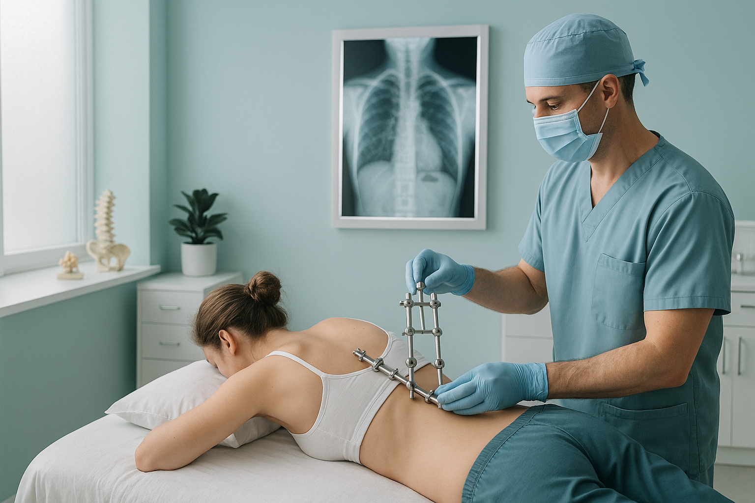
Other approaches include:
- Anterior spinal fusion. The surgeon reaches the spine from the front or side. This is less common but may be used for certain curve patterns.
- Vertebral body tethering (VBT). A flexible cord is attached to the outside of the curve to guide gradual correction. This keeps some spinal movement and is sometimes offered to younger adults or those with flexible curves.
- Osteotomy procedures. For stiff, severe adult curves, the surgeon may need to cut and reshape sections of bone to achieve correction before fusing.
Your surgeon will recommend the approach best suited to your curve type, flexibility and overall health.
What are the risks of scoliosis surgery?
Scoliosis surgery is a major operation, and all major surgery carries risk. Your surgical team will explain these in detail before you give consent. Common risks include infection, bleeding, nerve injury, hardware problems (such as a screw loosening) and the possibility that the fusion does not heal as expected (non-union).
There is also a small risk of spinal cord injury. This is why surgeons use intraoperative monitoring throughout the operation. In the longer term, fusing part of the spine means the segments above and below may take on more stress. This could cause new problems over the years.
Understanding the risks is an important part of making an informed decision. Your surgeon will weigh them against the likely benefits in your situation.
A note on MRI. EOS standing X-ray shows bones, joints and whole-body alignment beautifully. For soft tissue detail, such as discs, nerves or the spinal cord, MRI is usually the better tool. Many surgical plans need both. Your surgical team will advise which imaging you need.
What does recovery from scoliosis surgery look like?
Recovery from spinal fusion for scoliosis is gradual. Most people spend several days in hospital after the operation. Walking usually starts within a day or two, guided by the physiotherapy team.
Discharge from hospital, gentle walking, managing pain with medication. Avoid bending, lifting or twisting.
Walking distance increases gradually. Light daily activities start again. Most people are off strong painkillers by this stage.
Return to work (depending on the type of job) and gentle exercise. The fusion is still healing, so high-impact activity is avoided.
The fusion becomes solid. Most people return to their full range of daily activities. Follow-up imaging confirms healing and alignment.
Everyone recovers at a different pace. Your surgical team will give you a personal timeline and clear guidance on what you can do at each stage.
How does standing imaging help plan scoliosis surgery?
Standing, weight-bearing imaging shows your spine the way it actually behaves when you are upright. Lying-down scans relax the muscles. This can make a curve look different from how it appears in daily life. Gravity, posture and muscle tone all affect the shape and position of a scoliotic curve.
An EOS scan captures your entire skeleton from head to feet in one standing image, with about 90% less radiation than a standard X-ray, around ten times less. Your surgeon can see the full chain of alignment: how the curve affects your pelvis, hips and legs, not just the spine on its own.
Lying-down imaging (standard X-ray, CT or MRI) removes the effect of gravity. The curve may look smaller or different. Compensatory shifts in the pelvis or legs are hidden. This can give an incomplete picture for surgical planning.
Imaging before surgery with ScanAlign, standing EOS imaging gives surgeons measurable, weight-bearing data on coronal balance, sagittal balance and shoulder alignment. This information is critical for deciding how much correction to attempt and which levels to fuse.
- Surgical planning before an operation
- Monitoring curve progression over time
- Assessing whole-body alignment, not just the spine
- Follow-up after surgery with low radiation
- Detail on disc herniation or nerve compression
- Spinal cord assessment before complex surgery
- Soft tissue or inflammatory conditions
When should you seek urgent medical help?
If you have sudden numbness, weakness in your legs, or loss of bladder or bowel control, seek urgent medical attention. Contact your doctor straight away or go to your nearest emergency department. These symptoms can be a sign of serious nerve compression that needs immediate assessment, not a planned scan.
For symptoms that are not urgent, it is worth getting a detailed diagnosis before you start treatment or change the one you are on. Knowing whether the cause sits in the pelvis, the legs or the spine changes what will actually help.
Frequently asked questions about scoliosis surgery
Surgery is typically discussed when a curve is large and progressing, especially if it is affecting breathing or daily function. There is no single cut-off that applies to everyone. Your surgeon will look at the curve size alongside your symptoms, balance and overall health.
Yes. Adults with degenerative scoliosis, or with curves that have progressed since the teenage years, may be candidates. The goals and techniques can differ from surgery in teenagers, with more focus on pain relief, restoring balance and nerve decompression.
Spinal fusion joins two or more vertebrae together using rods, screws and bone graft. Over time, the fused section becomes one solid bone. This stabilises the curve and prevents further progression. It is the most commonly performed scoliosis surgery.
Most people are up and walking within a couple of days. A return to desk work often takes a few weeks, while more physical jobs may need several months. Full healing of the fusion usually takes six to twelve months.
Risks include infection, bleeding, nerve injury, hardware problems and non-union (failure of the fusion to heal). Serious complications such as spinal cord injury are rare. Your surgeon will explain all risks in detail before you give consent.
No referral is needed. ScanAlign is a private, self-pay clinic. You can book scoliosis imaging before surgery directly. Many people take their results to their surgeon or consultant afterwards.
An EOS scan uses about 90% less radiation than a standard X-ray, around ten times less. This makes it especially useful when repeated imaging is needed, such as monitoring a curve before or after surgery.
Yes. Standing imaging can show alignment problems that are hidden on lying-down scans. The curve may look different under gravity, and compensatory shifts in the pelvis or legs become visible. This can affect which levels are fused and how much correction is attempted.
The price for patients travelling from abroad is confirmed during the free video consultation. ScanAlign is a self-pay service.
Yes. In the UK, scoliosis surgery is available on the NHS for those who meet the criteria. Waiting times and access vary. Some people in the UK choose to have imaging before surgery done privately to speed up the planning process or to get a second opinion.
See your full alignment before surgery
A standing EOS scan gives your surgeon the weight-bearing detail they need for accurate planning. No referral needed.
Book a free video consultationSources
- Scoliosis Research Society. Patient information: surgical treatment of scoliosis. srs.org
- NHS. Scoliosis: treatment. nhs.uk/conditions/scoliosis/treatment
- Illés T, Somoskeöy S. The EOS imaging system and its uses in daily orthopaedic practice. International Orthopaedics, 2012;36(7):1405-1411.
Written by Abbas Dhami (Specialist Diagnostic Radiographer)
This page is general information and not a diagnosis.
ScanAlign is the trading name of CoreMed Solutions Ltd and operates under The Harley Street Hospital's CQC licence.