
Spine Anatomy: What Each Section of Your Back Actually Does
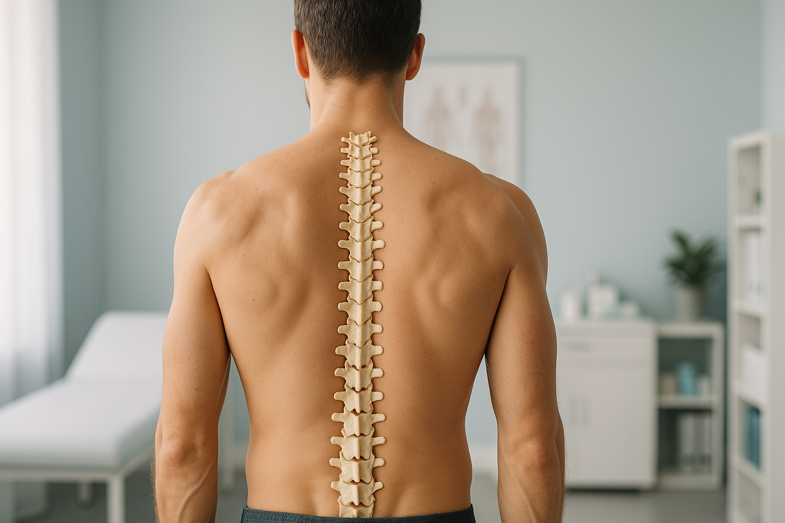
Your spine has five regions. They run from the cervical vertebrae in your neck down to the coccyx at the base. Each region has a different shape and a different job. The back muscles around these bones hold you upright, absorb load and allow movement. When one section is out of balance, other sections compensate. This often causes pain somewhere you would not expect.
Key points
- The spine has 33 vertebrae across five regions: cervical, thoracic, lumbar, sacrum and coccyx.
- Back muscles, including the erector spinae and multifidus, keep you stable and upright.
- The spine's natural curves are essential for shock absorption, but too much or too little curve can cause problems.
- Lying-down scans relax the muscles and can hide alignment issues that only appear when you are standing.
- A standing, weight-bearing scan shows how your whole spine works under gravity.
Understanding the anatomy of your spine is the first step to working out why your back, hip or knee hurts. Your body is a connected chain from your feet to your head. When one part of that chain is under strain, pain often shows up somewhere else entirely. This page goes through each region, the back muscles that support it, and why alignment matters.
What are the five regions of the spine?
The spine is divided into five separate sections. The vertebrae in each one are named by a letter and a number. From top to bottom, they are: cervical (C1 to C7), thoracic (T1 to T12), lumbar (L1 to L5), sacrum (five fused bones) and coccyx (three to four fused bones). That makes 33 vertebrae in total. Between most of the moveable ones sit intervertebral discs, which act as shock absorbers, and facet joints, which guide movement.
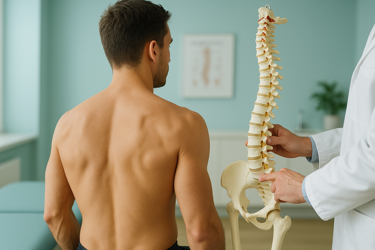
Supports the head and lets you look up, down and from side to side. This is the most mobile section.
Attaches to the ribs and protects the heart and lungs. It is less flexible and built for stability.
Carries most of your body weight. The lumbar spine has the largest vertebrae and is a common source of pain.
The sacrum connects the spine to the pelvis. The coccyx (tailbone) is a small anchor point for muscles and ligaments.
The spinal cord runs through a channel inside these vertebrae. Nerve roots branch out at each level. They send signals to your arms, legs and organs. A problem at any level can affect structures far away.
What do the back muscles do?
Your back muscles hold the spine upright, control movement and absorb everyday forces. Without them, the bones of the spine alone would buckle under the weight of your own body. There are two key groups worth knowing about.
A group of long muscles that run on either side of the spine, from the pelvis to the skull. They keep you upright and extend the back when you bend forward.
Small, deep muscles that span one to three vertebrae at a time. They fine-tune the position of individual segments and stabilise them under load.
The abdominal muscles, hip flexors and gluteal muscles work with the back muscles as a team. Weakness in any one group shifts extra load onto the others.
When one group of back muscles is weak or tight, it changes the way force travels through the spine. Over time, this can alter the natural curves. It can also put extra pressure on discs, facet joints or nerve roots. That is why pain in your lower back might actually start with weakness in your hip, or stiffness in your mid-back.
Why does the spine have natural curves?
The natural curves are there to absorb shock and spread load evenly. Seen from the side, a healthy spine has a gentle inward curve at the neck (cervical lordosis), an outward curve in the mid-back (thoracic kyphosis), and another inward curve at the lower back (lumbar lordosis). Together, these three curves make an S-shape that balances your head over your pelvis.
When these curves become too large or too flat, the body has to compensate. Too much forward rounding in the thoracic spine may cause the lumbar spine to increase its curve to keep you balanced. Too little lumbar curve may shift your head forward. You can read more about these patterns on our page about kyphosis and lordosis.
The way these curves relate to each other is called sagittal balance. When the curves of your spine are well matched, the muscles work efficiently and the joints wear evenly. When they are not, certain back muscles have to work harder than they should. This leads to fatigue, tightness and pain.
Why should spine alignment be checked while standing?
Because gravity is the force your spine manages every day, and lying down removes it. When you lie flat for a standard X-ray, CT scan or MRI, your muscles relax, your discs decompress, and your curves can change shape. The image may look reassuring. But it does not show how your spine actually behaves when you are on your feet.
A standing scan captures your spine under real-world load. Every vertebral body, every curve and every tilt is recorded exactly as it appears when gravity is acting on it. Your back muscles are active, your weight passes through your joints, and any misalignment or compensation can be seen.
At ScanAlign, unstitched full-body standing imaging captures the whole skeleton from head to ankle in a single upright exposure, so nothing is missed.
When you lie on a table, your spine flattens and your muscles switch off. A curve that causes pain under upright load may straighten out completely when you are lying flat. This means a lying-down scan might show "normal" anatomy while you are still struggling to stand comfortably.
Lying-down imaging is very useful for other purposes, but for alignment questions it can be misleading.
A full spine x-ray taken while standing gives your clinician the information they need to assess alignment across every region at once, not just one small section on its own.
When is MRI the better choice? EOS standing imaging shows bones, joints and alignment clearly. For soft-tissue problems such as disc bulges, nerve compression or spinal cord issues, MRI is usually the better tool. The two work together. They do not compete.
When should you get urgent help for back pain?
Most back pain is not an emergency. But some symptoms need immediate medical attention. If you have any of the following, contact your doctor, call your local emergency number, or go to the nearest emergency department. Do not wait to book a scan.
Sudden numbness, tingling or weakness in your legs needs urgent assessment.
Any loss of bladder or bowel control along with back pain is a medical emergency.
Intense pain that came on suddenly, especially after an injury or a fall, should be assessed promptly.
For persistent or unexplained back pain that has not responded to treatment, understanding your whole-body alignment can help your clinician find the underlying cause.
Before you start a treatment, or change the one you are on, it is worth getting a detailed diagnosis first. Knowing whether the cause sits in your pelvis, your legs or your spine changes what will actually help.
Would you like to understand how your spine alignment may be contributing to your symptoms?
Book a free video consultationFrequently asked questions about spine anatomy
There are 33 vertebrae in total: 7 cervical, 12 thoracic, 5 lumbar, 5 fused sacral and 3 to 4 fused coccygeal bones.
Cervical vertebrae are small and allow the most movement (your neck). Thoracic vertebrae attach to the ribs and provide stability. Lumbar vertebrae are the largest and carry the most weight. Each section has a different shape that suits its role.
The erector spinae muscles run along the full length of the spine and keep you upright. The multifidus muscles stabilise individual vertebrae. The core muscles, including the abdominal and hip muscles, work together with these to support spinal alignment.
Many scans are taken lying down. This relaxes the muscles and can hide alignment problems. A standing scan shows how your spine behaves under gravity, which may reveal issues that a lying-down scan misses.
An intervertebral disc sits between each pair of moveable vertebrae. It has a tough outer ring and a softer centre. Discs absorb shock, allow movement and help spread load evenly through the spine.
Facet joints are small paired joints at the back of each vertebra. They guide and limit movement, and stop the spine from twisting or bending too far. They can become a source of pain if they are overloaded or arthritic.
Yes. An EOS scan uses about 90% less radiation than a standard X-ray, around ten times less. It captures the full skeleton in one standing exposure without stitching multiple images together.
No. ScanAlign is a private, self-pay clinic. You can book a full spine standing scan directly, without a referral from your doctor or a consultant.
The price for patients travelling from abroad is confirmed during the free video consultation. This consultation can also help you decide whether a scan is the right next step for you.
Yes. A full-body standing scan captures everything from the base of the skull to the ankles. This lets a specialist see how the spine, hips and knees relate to one another, and find where compensation may be happening.
See how your spine really aligns
A free video consultation helps you understand whether a standing scan in London is the right next step for your back pain.
Book a free video consultationSources
- NHS. "Back pain." nhs.uk, 2023. Overview of causes, self-care and when to seek help.
- Illés, T., Somoskeöy, S. "The EOS imaging system and its uses in daily orthopaedic practice." International Orthopaedics, 2012; 36(7): 1405-1411.
- Panjabi, M.M. "The stabilizing system of the spine. Part I. Function, dysfunction, adaptation, and enhancement." Journal of Spinal Disorders, 1992; 5(4): 383-389.
Written by Abbas Dhami (Specialist Diagnostic Radiographer)
This page is general information and not a diagnosis.
ScanAlign is the trading name of CoreMed Solutions Ltd and operates under The Harley Street Hospital's CQC licence.