
How Does Your Skeleton Affect Your Squat and Deadlift Depth?
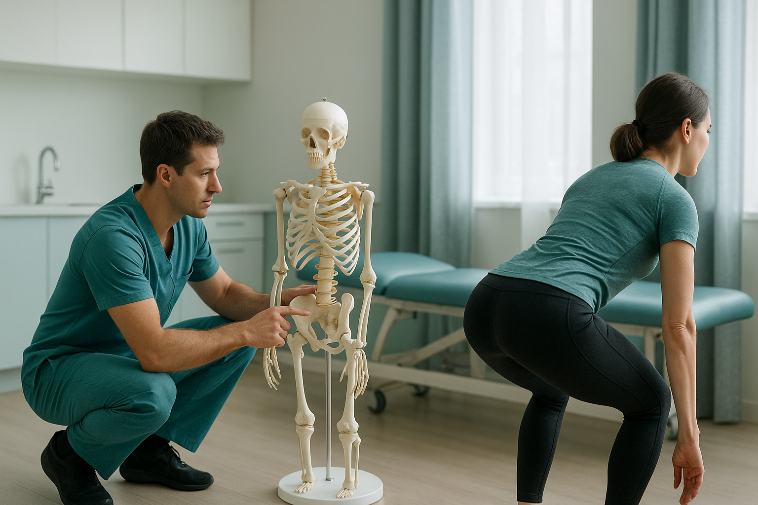
Your squat and deadlift form depends on your bone structure as much as your flexibility. Three things set hard limits on how deep you can go: the depth of your hip socket, the angle of your thigh bone, and the length of your torso compared with your femur. No amount of stretching changes the skeleton you were born with.
Key points
- Hip socket depth and direction vary hugely from person to person, and they decide how much range you have.
- Femoral neck angle and version (how the thigh bone twists) affect the best squat stance width for you.
- A longer femur compared with your torso forces you to lean further forward in the squat. This shifts stress to the lower back.
- "Butt wink" at the bottom of a squat is often a bony block, not a flexibility problem.
- A standing whole-body scan shows your real alignment under load. Imaging done lying down can miss this.
Why can't some people squat deep no matter how much they stretch?
Because their skeleton physically stops them. The hip joint is a ball and socket, but the size and angle of both parts are different in every person. When bone meets bone, no mobility drill can push you further. These three anatomical factors explain most limits in the squat and deadlift.
A deeper socket (acetabulum) covers more of the ball. This gives stability but blocks your range sooner. A shallower socket allows more movement before bone touches bone.
The neck of the thigh bone joins the shaft at an angle that differs from person to person. A steeper angle (coxa vara) and a flatter angle (coxa valga) each change how the femur moves inside the socket.
The thigh bone also has a natural twist. Hip anteversion and retroversion decide whether your knees naturally point inward or outward. So they also decide your most comfortable squat stance width.
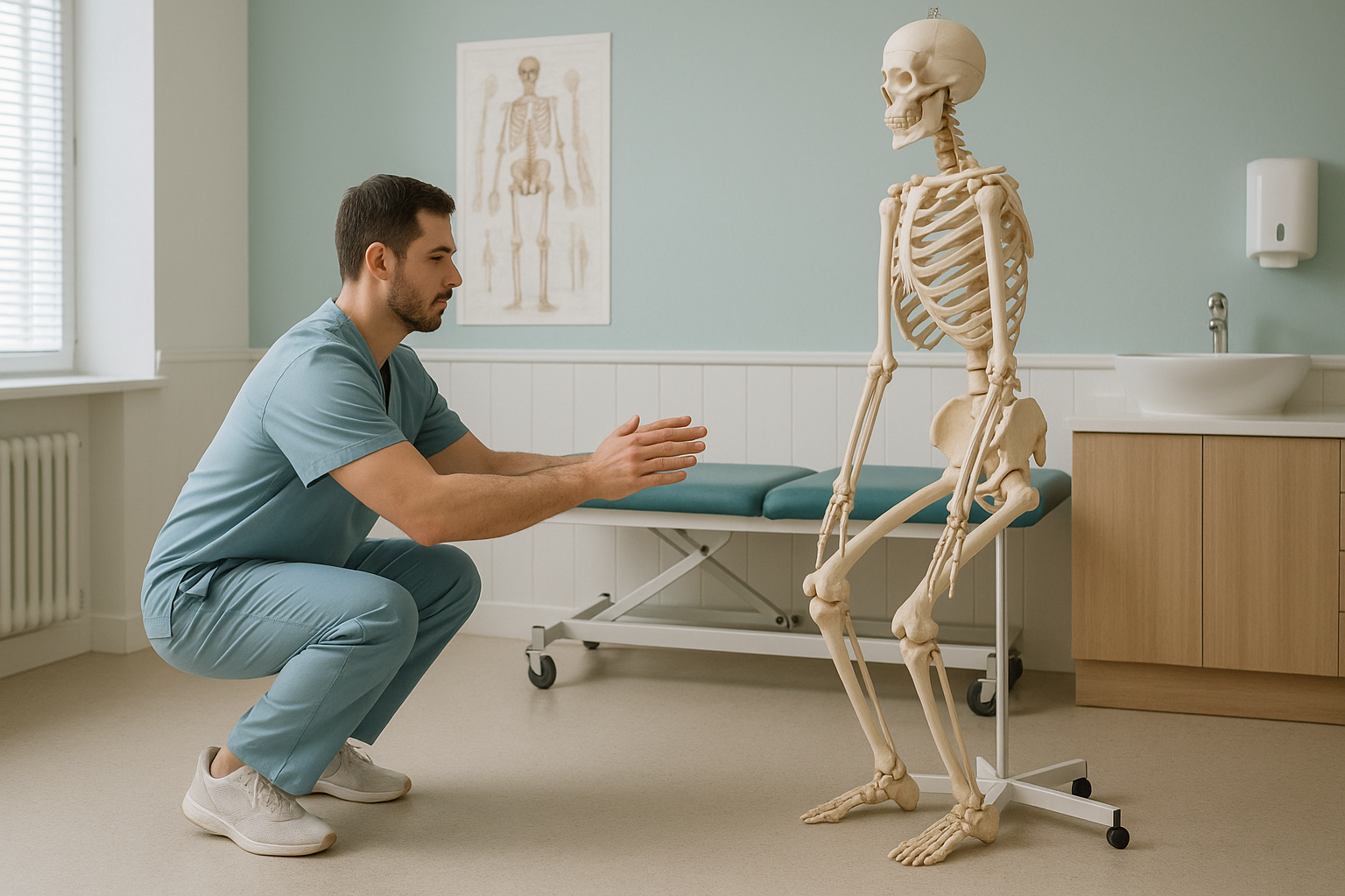
On top of these hip factors, the length of your limbs plays a big role. A longer femur compared with your torso forces you to lean further forward in a squat to keep the bar over your midfoot. That forward lean puts more load on the lower back. This is one common cause of back pain in the deadlift that has nothing to do with poor technique.
What are the signs that bone structure is limiting your squat or deadlift?
Limits from bone structure often look and feel different from tight muscles. These are the most common signs that your anatomy, not your mobility, is setting your limit.
Your pelvis tucks under at the bottom of the squat and your lower back rounds. This is often the femur pressing against the rim of the socket, not tight hamstrings.
A sharp, blocking feeling deep in the hip crease at the bottom of a squat. This can point to hip impingement (FAI), where extra bone on the socket or the head of the femur reduces the space in the joint.
One hip drops deeper than the other. Your left and right hip sockets are not always the same, so uneven depth is surprisingly common and simply part of your anatomy.
A persistent dull ache across the lower back, with no disc or nerve problem found. A short torso or an anterior pelvic tilt can shift load onto the spine during heavy pulls.
How does hip anatomy change your squat stance compared with your deadlift stance?
Squat stance width and toe angle are not the same for everyone. They depend on your acetabular orientation, the direction your hip socket faces. Someone whose socket faces more forward often squats most comfortably with a wider stance and toes turned out. Someone whose socket faces more to the side may prefer a narrower stance.
Back squat and front squat. The squat needs more hip flexion than any other barbell lift. Your hip socket depth, femoral version and torso-to-femur ratio all work together to set your natural bottom position. If your femur has significant anteversion, your knees may need to travel further forward and outward. Forcing them straight ahead can cause that familiar pinch at the front of the hip.
A muscle imbalance around the hips can make limits from your skeleton feel worse. It reduces the control you have through the range you do have.
Conventional and sumo deadlift. The deadlift uses less hip flexion than the squat, but your choice of stance matters just as much. Sumo deadlifts need a lot of external rotation and abduction. If your socket is deep or faces more forward, sumo may feel jammed at the start position. Conventional pulls, on the other hand, put more load on the lower back when your femur is long compared with your torso. Neither stance is wrong. The better one for you depends on your bones.
The key point is this: your skeleton is not a flaw. It is the fixed frame that your technique and training plan need to be built around. Forcing a stance or depth your bones will not allow leads to compensations further up or down the body, and in the end to pain or injury.
How is squat and deadlift form assessed with standing imaging?
A standing, weight-bearing scan shows your skeleton the way it really works: upright and under gravity. Imaging done lying down (standard X-ray, CT or MRI) relaxes the muscles and lets the joints settle into a different position. This can hide alignment problems that only appear when you are under load.
ScanAlign standing EOS imaging produces a full-length image from head to foot in a single exposure, using about 90% less radiation than a standard X-ray, around ten times less. From this image, a specialist can measure your femoral version, acetabular orientation, leg length differences and pelvic alignment, all while you are standing normally.
These measurements give you and your coach or physiotherapist objective information about your structure. Instead of guessing why your squat stops at a certain depth, you can see the reason in your bones. Then you can adjust your stance, shoe heel height or choice of exercises to match your anatomy.
When MRI is the better tool. EOS shows bones, joints and alignment. If you may have a labral tear, a disc problem or nerve compression, MRI is usually the better option because it shows soft tissue in detail. Your clinician can advise which scan is right for you.
Want to know whether your bones are shaping your squat or deadlift limits? A free video consultation from home can help you decide if a standing scan in London is the right next step.
Book a free video consultationWhat can you do if your skeleton limits your lifting depth?
Once you know which structures set the limit, you can work with that information instead of against it. These are general directions, not treatment advice. A qualified coach or clinician should guide the details.
- Adjust stance width and toe angle. Even small changes can open up the hip joint. This can let you reach more depth without forcing bone against bone.
- Choose the right deadlift style. Conventional, sumo and trap-bar deadlifts all load the hips differently. Your anatomy may strongly favour one over the others.
- Raise your heels. Weightlifting shoes or small heel wedges change your ankle and hip angles. This can make a real difference if you have long femurs.
- Accept your depth. Not every lifter needs to squat to full depth for strength or muscle growth (hypertrophy). Working within your available range, with control, is safer and often just as effective.
- Strengthen where you compensate. If your structure forces extra forward lean, building posterior chain strength helps you manage that lean safely.
If you think your structure includes a bony variation such as hip impingement (FAI) or significant hip anteversion and retroversion, a clear picture of your hip anatomy can save months of guesswork. You can book a hip and pelvis scan to see exactly what you are working with.
Before you start treatment, or change the one you are on, it is worth getting a detailed diagnosis. Knowing whether the cause sits in your pelvis, your legs or your spine changes what will actually help.
Frequently asked questions
If your hip socket is deep or your femur has limited version, bone meets bone before your muscles run out of stretch. No mobility drill changes the shape of a bone. Finding the structural limit lets you adjust your technique instead of chasing a depth your skeleton will not allow.
Butt wink is the rounding of the lower back at the bottom of a squat, caused by the pelvis tucking under. A small amount under light load is usually harmless. Under heavy load it increases stress on the lumbar spine. It is often a limit of your anatomy rather than a flexibility problem.
Yes. Your best stance width depends on your acetabular orientation and femoral version. A wider stance suits some hip structures, and a narrower one suits others. Trying different stances is useful, but imaging can show you exactly why one stance feels more natural.
It can contribute. If your hip sockets or femur proportions limit how far you can hinge, the lower back makes up the difference. This extra flexion under load can cause pain, even when there is no disc or nerve problem.
Neither is better for everyone. Sumo needs more hip abduction and external rotation, so it suits people with shallower sockets that face more forward. Conventional is often better for deeper sockets or sockets that face more to the side. Your anatomy should guide the choice.
A standing EOS scan captures your whole skeleton in one image while you are upright and bearing weight. This means alignment and load distribution are measured under real conditions. A standard X-ray usually covers one area at a time and is often taken lying down, which can hide alignment problems.
EOS uses about 90% less radiation than a standard X-ray, around ten times less. It is a very low-dose imaging method and is considered safe for adults, including those who may need follow-up scans over time.
No. ScanAlign is a private self-pay clinic, so you can refer yourself. You start with a free video consultation to check whether a scan is suitable for your situation.
The price for patients travelling from abroad is confirmed during the free video consultation, which will also confirm which scan package suits your needs. You can find more answers on the ScanAlign FAQs page.
Imaging can show your hip socket orientation, femoral version and limb proportions. These are the key factors that affect stance. Your coach or physiotherapist can then use that information to recommend a stance that fits your structure.
Stop guessing. See your skeleton.
A free video consultation takes a few minutes and helps you decide if a standing scan is the right next step for your training, before you plan a trip to London.
Book a free video consultationWritten by Abbas Dhami (Specialist Diagnostic Radiographer)
Sources
- Hartel, M.J. et al. (2016). "Determination of Femoral Neck Angle and Torsion." Journal of Orthopaedic Research, 34(8), 1460–1467.
- Mov, R. & Contreras, B. (2015). "Squatting Kinematics and Kinetics and Their Application to Exercise Performance." Journal of Strength and Conditioning Research, 29(12), 3497–3506.
- NHS. "Hip pain in adults." NHS.uk. Available at: nhs.uk/conditions/hip-pain.
This page is general information and not a diagnosis.
ScanAlign is the trading name of CoreMed Solutions Ltd and operates under The Harley Street Hospital's CQC licence.