

What Is a Vertebral Haemangioma, and Is It Dangerous?
A vertebral haemangioma is a benign (non-cancerous) collection of blood vessels inside a bone of the spine. It is one of the most common chance findings on spine scans, and it is almost always harmless. Most people never know they have one. The vast majority need no treatment at all.
Key takeaways
- Vertebral haemangiomas are found in up to 10% of spine scans and are almost always harmless.
- They are usually found by accident, when you have a scan for something else.
- The vast majority cause no symptoms and need no treatment.
- A very small number are classed as "aggressive" and may need a specialist to review them.
- If you have new numbness, weakness or bladder changes, get urgent medical help.
What exactly is a vertebral haemangioma?
A vertebral haemangioma is a small tangle of blood vessels that forms inside the body of a vertebra (one of the bones that make up your spine). It is a benign vascular lesion. This means it is not cancer, and it does not spread to other parts of the body.
Think of it as a tiny cluster of extra blood vessels woven into the spongy inside of a spinal bone. They develop slowly, often over many years. In most people they stay small and completely silent.
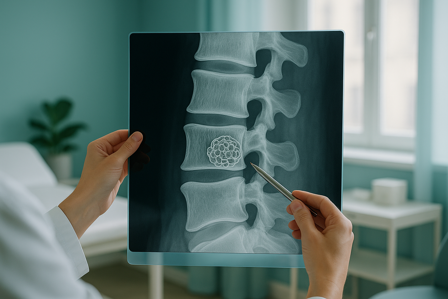
Vertebral haemangiomas are very common. Studies suggest they show up in roughly 10% of spine MRI scans. Most are picked up as an incidental finding. This means the scan was done for a completely different reason, such as checking back pain, and the haemangioma was spotted along the way.
Does a vertebral haemangioma cause symptoms?
In the vast majority of cases, no. Most vertebral haemangiomas cause no symptoms at all. You cannot feel them, and they do not cause pain.
The overwhelming majority of haemangiomas cause no pain at all. If your scan report mentions one, it is very likely unrelated to the symptoms your scan was done for.
They are usually noted on a scan report as a side observation. Your radiologist may describe it as "an incidental finding of no clinical significance."
In very rare cases, a large or "aggressive" haemangioma may grow into the spinal canal and press on nerves. This can cause pain in one area, tingling or weakness.
Your scan report may mention a vertebral haemangioma. If you have no new nerve symptoms (numbness, weakness, bladder or bowel changes), you can almost certainly feel reassured by the finding. It is overwhelmingly likely to be harmless.
What is the difference between a typical and an aggressive haemangioma?
Radiologists generally put vertebral haemangiomas into two groups, based on how they look on scans. This matters because it guides whether any follow-up is needed.
- Small, contained within the vertebral body
- Has a typical "polka dot" look on scans
- Causes no symptoms at all
- Needs no treatment and no follow-up
- Makes up the vast majority of cases
- Larger and may extend beyond the bone
- Can press on the spinal cord or nerve roots
- May cause pain, numbness or weakness
- Usually needs assessment by a specialist
- Makes up a very small fraction of all haemangiomas
Your scan report will usually describe the appearance in enough detail for your doctor to tell whether it is typical or aggressive. If you are not sure, ask your doctor or the radiologist who wrote the report to explain.
How is a vertebral haemangioma found?
Almost all vertebral haemangiomas are found by chance on a spine MRI. MRI is very good at picking them up, because of the way the blood vessels inside the lesion look on different scan sequences. They can also be seen on CT scans, and sometimes on a full spine x-ray, but MRI gives the most detail.
A note on types of scan. MRI is excellent for finding and describing soft-tissue structures, including haemangiomas, discs and nerves. EOS and standing x-rays, such as those offered by ScanAlign standing EOS imaging, are designed to show bones, joints and whole-body alignment under gravity. Each type of scan answers a different clinical question, and they work well together.
A haemangioma sits inside the bone, so it does not affect your posture or alignment. If you had a scan for back pain and a haemangioma was noted, your clinician will usually focus on the other findings (such as disc or alignment problems) as the more likely explanation for your symptoms.
Does a vertebral haemangioma need treatment?
For most people, the short answer is no. A typical vertebral haemangioma with no symptoms is a benign spine lesion. It does not need any treatment, monitoring or lifestyle changes. You can exercise, lift, stretch and carry on with daily life as normal.
There is no evidence that a typical haemangioma will grow, turn into cancer or cause problems in the future. Most clinicians will not even mention it at follow-up appointments, because it is seen as a normal variant.
In the very rare case where a haemangioma is classed as aggressive and is pressing on nerves and causing symptoms, a specialist may consider treatment. This would always be managed by a spinal or neurosurgical team, not by a diagnostic imaging clinic. If you are unsure whether your findings mean you should see a specialist, read our guide on when to see a specialist.
When should you worry about a spinal haemangioma?
For the vast majority of people, there is no need to worry at all. A typical vertebral haemangioma is one of the most benign findings possible on a spine scan. It is not a tumour that will spread, and it will not weaken your spine.
However, get urgent medical help (your doctor or the emergency department of your nearest hospital) if you develop any of the following, whatever your scan shows:
- New or worsening numbness or tingling in your legs or arms
- Weakness in your legs that makes walking difficult
- Loss of bladder or bowel control
- Sudden, severe back pain that is very different from your usual symptoms
These symptoms can have many causes and are not specific to haemangiomas. They always need a prompt medical check.
Before you start treatment for back pain, or change the one you are on, it is worth getting a detailed diagnosis. Knowing whether the cause sits in the pelvis, the legs or the spine changes what will actually help.
If your back pain is ongoing and earlier scans have not given clear answers, a standing assessment can sometimes show alignment problems that lying-down scans miss. Speak to our team to find out whether it could help you.
Book a free video consultationFrequently asked questions about vertebral haemangiomas
No. A vertebral haemangioma is a benign (non-cancerous) collection of blood vessels. It does not spread, and it is not related to bone cancer or metastatic disease.
Typical haemangiomas do not cause pain. If you have back pain and a haemangioma was found by chance on your scan, the pain is almost certainly coming from something else, such as a disc, joint or alignment problem.
Very common. Studies suggest they appear in roughly 10% of all spine MRI scans. Many more people probably have them without ever having a scan.
For a typical haemangioma with no symptoms, no follow-up is needed. If the radiologist describes it as atypical or aggressive, your doctor may advise more scans or a specialist opinion.
Most stay the same for life. In rare cases an aggressive haemangioma may slowly get bigger. This is uncommon, and it would usually be picked up on routine scans if you are being monitored for another reason.
No. A typical haemangioma does not affect the strength of your spine. You can exercise, lift weights and stay active as you normally would.
On MRI, a typical haemangioma looks bright on certain sequences, because of the fat and blood vessels it contains. On CT, it often shows a typical "polka dot" pattern caused by thickened bone struts. These appearances are distinctive enough that more tests are rarely needed.
Yes. It is not unusual to have haemangiomas in more than one vertebra. Several typical haemangiomas are still considered benign and generally need no treatment.
EOS imaging is designed to assess bones, joints and whole-body alignment, not the inner structure of single vertebrae. MRI is the best tool for finding and describing haemangiomas. If you also have concerns about alignment or posture, a standing scan can add useful information.
At ScanAlign, you do not need a referral from your doctor. You can book a standing spine scan directly. For MRI, most private providers also accept self-referrals, though some may prefer a letter from your doctor.
Have questions about your spine scan findings?
If earlier scans have not fully explained your back pain, a free video consultation can help you understand whether a standing alignment assessment is right for you.
Book a free video consultationWritten by
Abbas Dhami (Specialist Diagnostic Radiographer)
Sources
- Rodallec MH, Feydy A, Larousserie F, et al. Diagnostic imaging of solitary tumors of the spine: what to do and say. RadioGraphics. 2008;28(4):1019-1041.
- Murphey MD, Fairbairn KJ, Parman LM, et al. Musculoskeletal angiomatous lesions: radiologic-pathologic correlation. RadioGraphics. 1995;15(4):893-917.
- NHS. Back pain: causes, treatment and when to see a GP. nhs.uk/conditions/back-pain.
This page is general information and not a diagnosis.
ScanAlign is the trading name of CoreMed Solutions Ltd and operates under The Harley Street Hospital's CQC licence.