
What Does a Weight-Bearing Foot X-ray Show That a Lying Scan Misses?
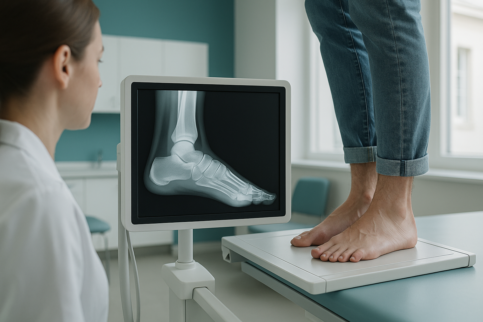
A weight-bearing foot X-ray takes a picture of your foot while it carries your full body weight. It shows arch collapse, true joint spacing and hindfoot alignment. These disappear the moment you lie down. Gravity and load shape the way your bones sit. So a standing image gives your clinician the real picture that a normal lying X-ray cannot.
Key takeaways
- Standing puts your full body weight on your foot. This shows how your arch, joints and heel truly sit.
- Lying scans relax the muscles and let bones drift into an unloaded position. This can hide real problems.
- EOS standing imaging uses about 90% less radiation than a standard X-ray, around ten times less.
- You do not need a referral from your doctor. You can book a private foot X-ray in London yourself.
- A specialist reports on your images. They can be shared with your podiatrist or orthopaedic surgeon.
What does a standing foot X-ray actually show?
A standing foot X-ray shows the true relationship between every bone in your foot and ankle while they are loaded. When you stand, your body weight presses the joints together and stresses the ligaments. It does this exactly as it does with every step you take. The image captures arch height, the angle between your heel bone and your forefoot, and the spacing between each joint. All of these are shown in their working, real-life position.
These details matter. Podiatrists and orthopaedic foot surgeons rely on loaded views to plan orthotics, injections or surgery. A non-weight-bearing image may look normal, even when the foot behaves very differently under load.
Why does a lying scan miss foot problems?
Without gravity, the muscles relax, the arch lifts and the joints spring open. That is why a lying scan misses foot problems. The image shows an ideal skeleton, not the one you actually walk on. This is the basic difference between weight-bearing versus lying imaging.
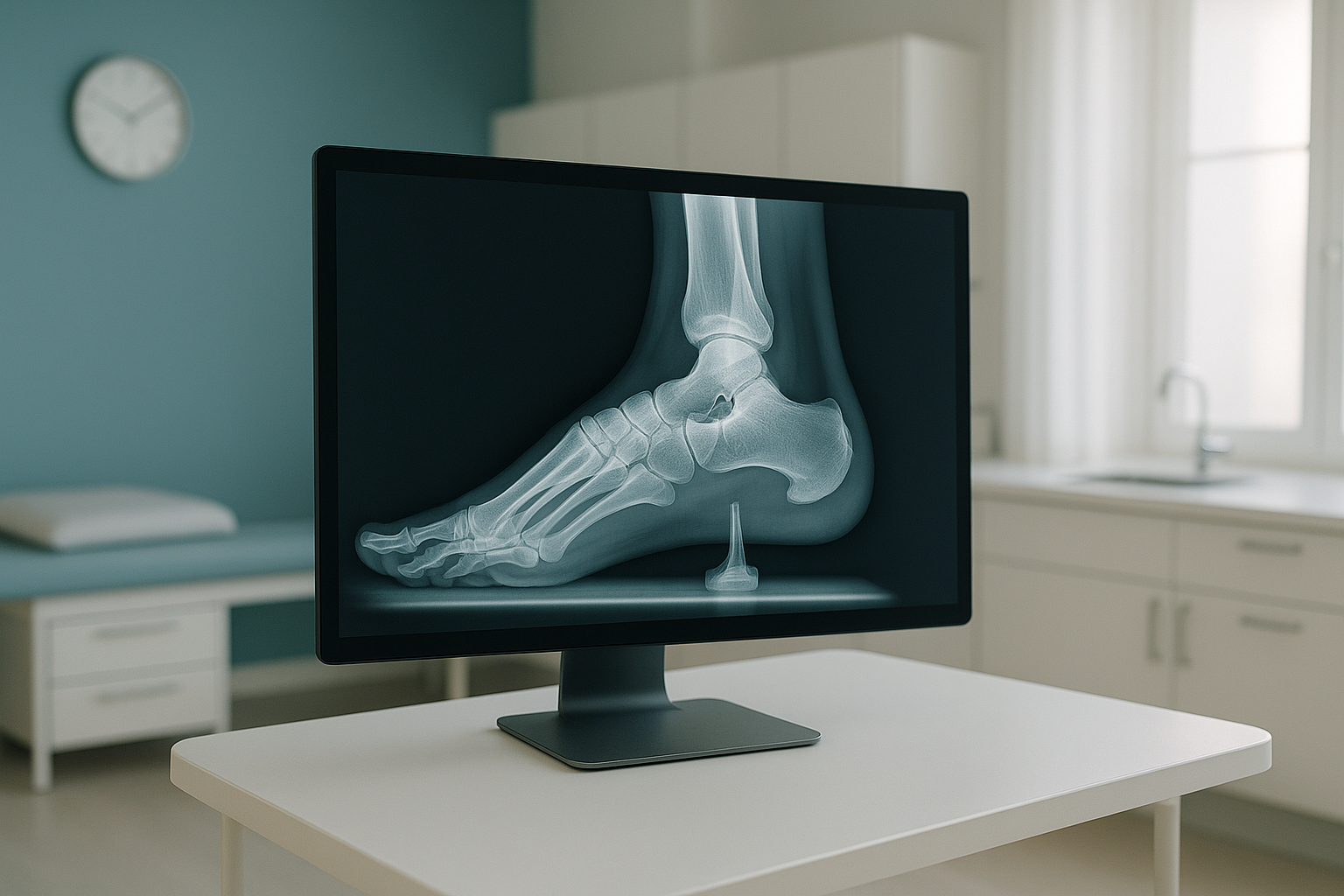
Your body is a connected chain from your feet to your head. A collapsed arch can shift your ankle inwards and change the angle of your knee. It can also create strain in your hip or lower back. A standing weight-bearing X-ray captures that chain reaction. A lying scan cannot. To learn more about how the two compare, read our guide to weight-bearing or traditional imaging.
What is included in a weight-bearing foot scan at ScanAlign?
Your appointment at ScanAlign, standing EOS imaging in London includes everything you need for a complete foot alignment assessment.
| Feature | Standard lying X-ray | EOS standing scan |
|---|---|---|
| Position | Lying on a table | Standing, full weight on both feet |
| Radiation | Standard dose | About 90% less, around ten times less |
| Image scale | Magnified (varies with distance) | Non-magnified, true 1:1 size |
| Alignment data | Limited (unloaded bones) | Full loaded alignment of foot and ankle |
| Report | Varies by provider | Specialist radiographer report included |
EOS produces a non-magnified image at true 1:1 scale. So measurements taken from your scan reflect the real size of your bones and joints. This is especially useful for surgical planning, where even small measurement errors can affect the outcome.
The price for patients travelling from abroad is confirmed during the free video consultation. You can also visit the EOS scan cost page.
When MRI is the better choice: EOS is excellent at showing bones, joints and alignment. Your clinician may suspect a soft-tissue problem, such as a torn tendon, plantar fascia damage or nerve compression. In that case, MRI is usually the better tool. We are always happy to advise during your free consultation.
How do I book a private weight-bearing foot X-ray in London?
You can book yourself, without a letter from your doctor. Here is how it works.
A short call with our specialist radiographer. We listen to your symptoms, confirm that a weight-bearing foot X-ray is the right scan for you and answer any questions.
You visit us at 19 Harley Street, London. You stand naturally in the EOS system while it captures your foot and ankle under load. The scan takes only a few seconds.
A specialist reviews and reports on your images. The report and images are shared with you. If you wish, they are also sent directly to your podiatrist or orthopaedic surgeon.
With accurate, loaded images, your treating clinician can plan the most effective next step. This may be orthotics, physiotherapy or a surgical opinion.
Who should consider a weight-bearing foot and ankle X-ray?
A standing foot scan is useful for anyone whose foot or ankle symptoms change with activity or load. It is especially relevant in the following situations.
- Flat feet or fallen arches that get worse on standing
- Bunion or forefoot deformity assessment
- Ankle pain or instability under load
- Planning before foot or ankle surgery
- Ongoing foot pain despite normal lying X-rays
- Suspected stress fracture (MRI or bone scan may be better)
- Soft-tissue injury such as a torn Achilles tendon
- Unable to stand without support for a few seconds
- Acute injury that needs urgent emergency assessment
If you are unsure whether a weight-bearing scan is right for you, our free video consultation is the easiest way to find out. There is no obligation.
Before you start a treatment, or change the one you are on, it is worth getting a detailed diagnosis. Knowing whether the cause sits in the pelvis, the legs or the spine changes what will actually help.
Ready to see your foot under real load? Our specialist radiographer can explain what a standing scan would show in your own situation.
Book a free video consultationFrequently asked questions about weight-bearing foot X-rays
It is an X-ray taken while you stand on your feet. Your bones, joints and arches are captured under your full body weight. This shows how your foot actually works, unlike a lying X-ray where gravity is removed.
When you lie down, muscles relax and bones drift into an unloaded position. Problems like arch collapse, joint narrowing and heel tilt only appear under load. A standing scan captures these real-life changes.
No. You can book yourself at ScanAlign. We offer a free video consultation first to make sure the scan is right for your symptoms.
EOS uses about 90% less radiation than a standard X-ray, around ten times less. This makes it one of the lowest-dose options available for skeletal imaging.
The scan itself takes only a few seconds. The whole appointment, including positioning and any questions, is a short visit.
Yes. We can share your images and report directly with any treating clinician you choose. This may be a podiatrist, orthopaedic foot surgeon or physiotherapist.
EOS captures images while you stand. It uses much less radiation and produces non-magnified, true 1:1 scale images. A standard X-ray is usually taken lying down, with a higher dose and some image magnification.
It is one of the most reliable ways to assess flat feet. It shows exactly how much your arch drops when you stand. Lying scans often make a flat foot look normal, because the arch lifts without load.
The price for patients travelling from abroad is confirmed during the free video consultation. You can also visit our EOS scan cost page.
Yes. EOS uses about 90% less radiation than a standard X-ray. So it is well suited to follow-up imaging when your clinician needs to check for changes over time.
See your feet the way they really work
A short, free video call with our specialist radiographer will help you understand whether a standing scan is the right next step for your foot or ankle concern.
Book a free video consultationWritten by Abbas Dhami (Specialist Diagnostic Radiographer)
Sources
- Defined, S. et al. "Weight-bearing CT compared with conventional radiography for the assessment of hindfoot alignment." Foot & Ankle International, American Orthopaedic Foot & Ankle Society.
- NHS guidance on foot problems and when to seek imaging. NHS.uk.
- Deschênes, S. et al. "EOS low-dose radiography system: a systematic review of clinical applications." European Journal of Radiology, 2010.
This page is general information and not a diagnosis.
ScanAlign is the trading name of CoreMed Solutions Ltd and operates under The Harley Street Hospital's CQC licence.