
What Can an MRI Not Show You?
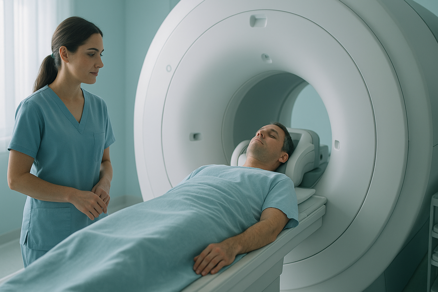
An MRI is excellent at showing soft tissue, such as discs, ligaments and nerves. But it cannot show how your skeleton lines up under your own body weight, because you lie down inside the scanner. So posture problems, joint shifts under weight and whole-body compensation patterns cannot be seen on a standard MRI.
Key takeaways
- An MRI shows soft tissue very well, but it cannot assess how the bones line up under load.
- Lying down relaxes the muscles and joints. This can hide problems that only show up under upright load.
- A "normal" MRI does not mean nothing is wrong. It may mean the wrong question was asked.
- Standing, weight-bearing imaging captures the whole skeleton, from head to feet, in one view.
- Many people with ongoing pain benefit from both an MRI and a standing alignment assessment.
What does an MRI miss?
An MRI misses anything that depends on gravity, posture or the load of your body weight. It is superb at showing soft tissue detail, but it has clear blind spots. Here are the main ones.
Why does lying down for a scan matter?
Lying down takes gravity out of the picture. Your muscles relax, your discs decompress, and your joints settle into a position they are never really in during daily life. As a result, an MRI taken lying down can look reassuringly normal, even when something is clearly wrong when you are upright.
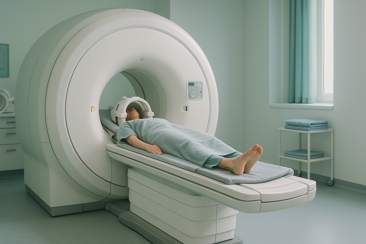
This is one of the key differences between weight-bearing and lying-down imaging. A condition like spinal stenosis, for example, may narrow the spinal canal more when you are upright and gravity presses down on the spine. Lying down can make that narrowing look less significant than it really is.
Why is my MRI normal but I am still in pain?
A normal MRI does not mean nothing is wrong. It means nothing was found in the soft tissue of the area that was scanned, while you were lying down. Several things could still be going on.
Research shows that many people with no back pain at all have disc bulges that can be seen on MRI. A finding does not always explain the pain, and a clear scan does not always rule it out. You can read more about when an MRI shows nothing but the pain remains.
How does the body compensate and hide the real problem?
Your body is a connected chain from your feet to your head. When one part is out of line, other parts shift to keep you balanced. This compensation can move the pain far away from its actual cause.
For example, a slight tilt in your pelvis can change how your lower back curves. That in turn can shift your mid-back and even the position of your neck. An MRI of your neck may look entirely normal, because the problem started lower down, in a part of the body that was never scanned.
Only a whole-body image, taken while you stand and carry your own weight, can reveal these compensation patterns in a single view. That is the whole-body picture that imaging of a single area misses.
Want to see how your whole body lines up, not just one section? A standing scan shows the full picture.
Book a free video consultationWhat does a standing whole-body scan show that an MRI cannot?
A standing scan captures your entire skeleton, from your head to your feet, in one image. Gravity acts on your body exactly as it does in daily life. It shows how every joint lines up under real-world load.
At ScanAlign, whole-body standing imaging uses EOS technology, which takes a full-length standing X-ray with about 90% less radiation than a standard X-ray, around ten times less. The image lets a specialist see alignment, compensation and changes in joint space that lying-down scans simply cannot capture.
When is an MRI still the right choice? An MRI is usually the better tool when your clinician needs to see soft tissue such as discs, ligaments, cartilage or nerves. EOS shows bones, joints and alignment. The two scans answer different questions, and many people benefit from having both. Neither replaces the other.
- Ongoing back, hip or knee pain with a normal MRI
- Pain that gets worse when you stand or walk
- A feeling that one side of your body works differently
- Suspected alignment or posture problems
- Planning before hip or knee replacement surgery
- Sudden numbness or weakness in your legs
- Loss of bladder or bowel control
- Sudden, severe pain after an injury
- Contact your doctor, or go to your nearest emergency department
Before you start a treatment, or change the one you are on, it is worth getting a detailed diagnosis. Knowing whether the cause sits in your pelvis, your legs or your spine changes what will actually help.
Frequently asked questions
Yes. An MRI is excellent for soft tissue, but it scans one area at a time while you lie down. Problems linked to posture, alignment or the load of your body weight can be invisible on an MRI, because gravity is not acting on the body.
A clear MRI means no soft tissue problem was found in that area, while you were lying down. Your pain may have a posture or alignment cause that only shows when you are upright and carrying your weight.
An MRI uses magnets to image soft tissue while you lie flat. A standing X-ray, such as EOS, images your bones and joints while you stand. It captures how your skeleton lines up under gravity. They answer different questions and often work well together.
No. Posture involves the whole body carrying its weight. Because an MRI scans you lying down and images one section at a time, it cannot assess how your posture works when you are standing.
EOS is a low-dose standing X-ray system that captures your full skeleton, from head to feet, in a single image. It uses about 90% less radiation than a standard X-ray, around ten times less. It is designed to show alignment and the shape of the joints under natural weight-bearing conditions.
EOS uses about 90% less radiation than a standard X-ray, around ten times less. It is one of the lowest-dose skeletal imaging methods available.
No. ScanAlign accepts self-referrals. You start with a free video consultation to check that a standing scan is right for your situation.
The price for patients travelling from abroad is confirmed during the free video consultation.
Absolutely. Many people benefit from both. An MRI shows soft tissue detail, such as discs and nerves. A standing EOS scan shows how the bones line up and the shape of the joints under load. Together they give a more complete picture.
Think about whether a weight-bearing assessment might reveal what the MRI could not. A free video consultation with ScanAlign can help you work out whether a standing scan is a useful next step. You can book a standing whole-body scan without a referral.
Still in pain after a normal MRI?
A free video consultation, from wherever you are, helps you find out whether a standing whole-body scan could reveal what lying-down imaging missed.
Book a free video consultationSources
- Brinjikji W, Luetmer PH, Comstock B, et al. Systematic literature review of imaging features of spinal degeneration in asymptomatic populations. AJNR Am J Neuroradiol. 2015;36(4):811-816.
- Dubousset J, Charpak G, Dorion I, et al. A new 2D and 3D imaging approach to musculoskeletal physiology and pathology with low-dose radiation and the standing position: the EOS system. Bull Acad Natl Med. 2005;189(2):287-297.
- National Institute for Health and Care Excellence (NICE). Low back pain and sciatica in over 16s: assessment and management. NICE guideline [NG59]. 2016 (updated 2020).
Written by Abbas Dhami (Specialist Diagnostic Radiographer)
This page is general information and not a diagnosis.
ScanAlign is the trading name of CoreMed Solutions Ltd and operates under The Harley Street Hospital's CQC licence.