
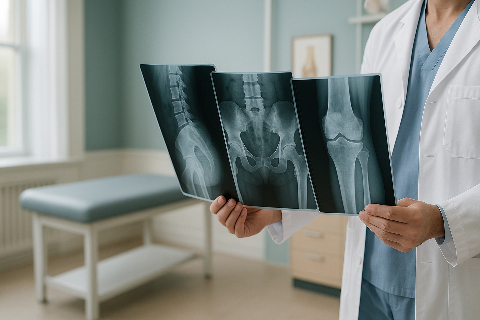
Which Scan Do You Need for Back, Hip or Knee Pain?
The right scan depends on the clinical question, not just where it hurts. X-rays show bones. MRI shows soft tissue such as discs and ligaments. CT gives detailed cross-sections of bone. An EOS standing scan shows the alignment of your whole body under load, with about 90% less radiation than a standard X-ray, around ten times less. Start with the question your clinician needs answered. Then choose the test that answers it.
Key takeaways
- Each type of scan is good at something different. No single scan answers every question.
- Bones and alignment need X-ray or EOS. Soft tissue, discs and nerves need MRI.
- Scans done lying down can hide alignment problems that only appear when you stand.
- You do not always need a referral from your doctor. A free video consultation can help you decide.
- For details on cost, safety and what to expect, see our frequently asked questions.
If you have had pain for weeks or months and are not sure which scan to ask for, you are not alone. There are several types of medical imaging. Each one is made for a different job. Choosing the wrong one can mean lost time, radiation you did not need, or a result that still does not explain your symptoms.
This page explains the most common scans in plain words. It helps you work out which one fits your situation.
A note on MRI. EOS shows bones, joints and alignment very clearly. For soft tissue, discs or nerves, MRI is usually the better tool. Often people need both. If you are unsure, a consultation can help you decide.
What are the main types of medical scans for pain?
Six common imaging tools are used for musculoskeletal problems. Each one is best at a specific task.
Should I get an X-ray, MRI or CT for my pain?
It depends on what your clinician needs to see. The table below is a quick guide. For a closer look at how EOS compares, read about EOS compared with X-ray, CT and MRI.
| Scan | Best for | Position | Radiation | Soft tissue? |
|---|---|---|---|---|
| X-ray | Fractures, arthritis, bone spurs | Lying or standing (one area) | Low | No |
| MRI | Discs, ligaments, nerves, cartilage | Lying down | None | Yes |
| CT | Complex fractures, 3-D bone detail | Lying down | Higher | Limited |
| EOS | Whole-body alignment, leg length, planning before surgery | Standing | Very low (about 90% less than X-ray) | No |
| Ultrasound | Tendons, bursae, guided injections | Various | None | Yes (superficial) |
| DEXA | Bone density / osteoporosis screening | Lying down | Very low | No |
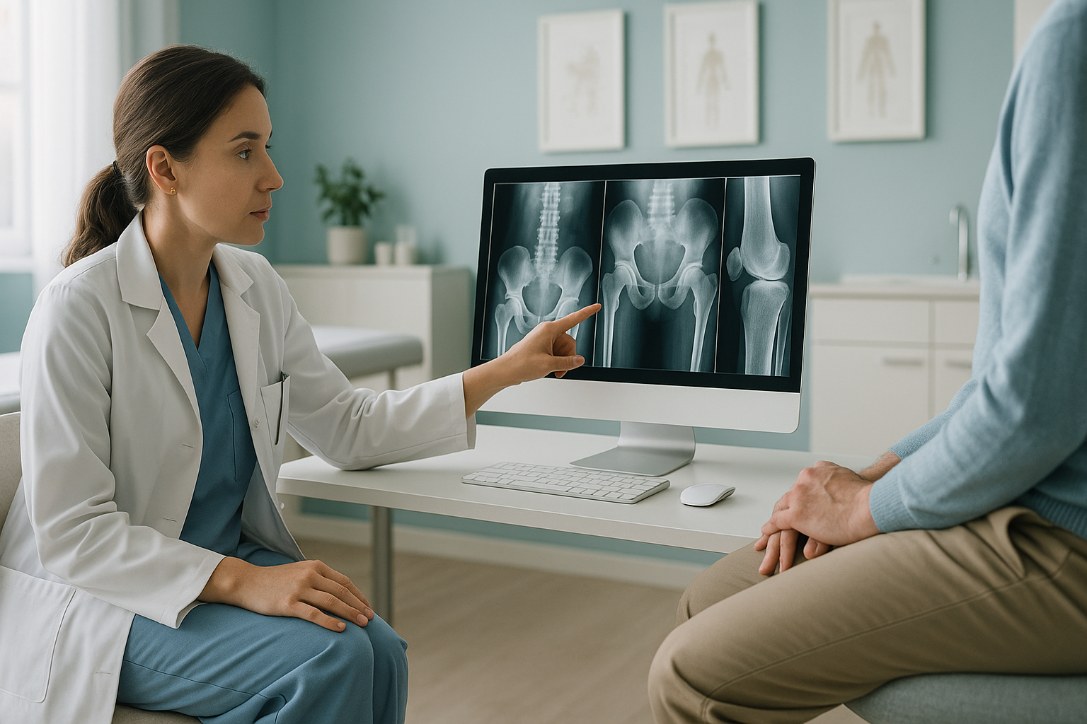
Why does standing up during a scan matter?
Standing up puts your full body weight on your skeleton. That changes everything. When you lie down for a standard X-ray, CT or MRI, gravity is taken away and your muscles relax. A tilted pelvis may look level. A curved spine may straighten. A knee that tracks poorly under load may look normal.
Your body works as a connected chain from your feet to your head. A problem in one area, such as a slight difference in leg length or a shifted pelvis, can force another area to compensate. That compensation often shows up as pain somewhere else entirely. A standing, whole-body EOS scan captures this full picture in a single image. So the real cause of your symptoms is far less likely to be missed.
To see how this applies to specific areas, visit our back pain clinic page or our guide to hip pain and alignment.
Not sure whether a standing alignment scan is right for you? A short video call with our team can help you check which scan suits you, before you plan any travel to London.
Book a free video consultationHow do I decide which scan I actually need?
Start with the clinical question, not the machine. Here is a simple way to think about it.
Define the question
Is the problem with the bone (fracture, arthritis, alignment) or with soft tissue (disc, ligament, nerve)? The answer narrows your options straight away.
Consider position
Does your pain get worse when you stand or walk? If so, a scan done lying down may miss what a standing scan would show.
Think about the whole chain
If a scan of one area has already come back "normal" but you still have symptoms, a view of whole-body alignment might reveal a compensating problem somewhere else.
Ask a professional
A radiographer or clinician can match the right scan to your situation. At ScanAlign, private standing imaging in London, we offer a free video consultation to help you work this out before you commit.
- Long-standing back, hip or knee pain with no clear diagnosis
- Pain that gets worse when standing or walking
- A suspected difference in leg length or a pelvic tilt
- Questions about alignment before or after surgery
- You suspect a disc bulge, nerve compression or ligament tear (MRI is usually better)
- You need a complex fracture mapped in 3-D (CT is usually needed)
- You are checking bone density (a DEXA scan is the right tool)
Before you start a treatment, or change the one you are on, it is worth getting a detailed diagnosis. Knowing whether the cause sits in the pelvis, the legs or the spine changes what will actually help.
Red-flag symptoms. If you have sudden numbness, weakness, or loss of bladder or bowel control along with back or leg pain, do not wait for any scan. Contact your doctor or go to the nearest emergency department straight away.
Frequently asked questions about choosing a scan
It depends on the suspected cause. If the concern is arthritis, alignment or a fracture, an X-ray or a standing EOS image is a good first step. If an injury to a ligament, meniscus or cartilage is suspected, MRI is usually more suitable because it shows soft tissue in detail.
For disc or nerve problems, MRI is generally the first choice. For spinal alignment, scoliosis or back pain linked to posture, a standing X-ray or EOS scan shows more because it captures your spine under load. Many people benefit from both.
An X-ray can show arthritis and narrowing of the joint space. MRI is better for labral tears and early cartilage damage. If your clinician suspects the pain is linked to pelvic tilt or alignment, a standing EOS scan shows how the hip sits in relation to the whole skeleton.
Neither is better in every case. CT is excellent for detailed bone imaging and is quicker to do. MRI is excellent for soft tissue. The right choice depends entirely on the clinical question being asked.
An EOS scan takes a full-length, standing X-ray from your head to your feet in a single pass. It uses about 90% less radiation than a standard X-ray, around ten times less. A regular X-ray captures one area. EOS shows how your entire skeleton lines up under your own body weight.
No. ScanAlign is a private, self-pay clinic. You can refer yourself. We recommend starting with a free video consultation so we can confirm whether an EOS scan is the right choice for your situation.
An EOS scan uses about 90% less radiation than a standard X-ray, around ten times less. This makes it one of the lowest-dose options available for imaging the skeleton.
An EOS scan images you from head to feet in one standing pass. This gives a complete view of skeletal alignment. Standard X-rays, MRI and CT usually focus on one area at a time.
For patients travelling from abroad, the price is confirmed during the free video consultation. You can also visit our frequently asked questions page.
You stand still inside the scanner for around 15 to 20 seconds while the image is taken. There is no enclosed tube and no loud noise. You can wear normal clothes without metal fastenings. Full details are on our frequently asked questions page.
Still not sure which scan is right for you?
A short video call with our team can help you work out the best next step, with no obligation.
Book a free video consultationAuthor: Abbas Dhami (Specialist Diagnostic Radiographer)
Sources
- NHS. "X-ray." nhs.uk. Accessed 2024.
- Royal College of Radiologists. iRefer: Making the Best Use of Clinical Radiology. RCR guidelines for imaging referral.
- Deschênes S, Charron G, Beaudoin G, et al. "Diagnostic imaging of spinal deformities: reducing patients' radiation dose with a new slot-scanning X-ray imager." Spine. 2010;35(9):989-994.
This page is general information and not a diagnosis.
ScanAlign is the trading name of CoreMed Solutions Ltd and operates under The Harley Street Hospital's CQC licence.