
What Is Blount's Disease, and When Do Bow Legs Need Treatment?
Blount's disease is a growth plate disorder at the top of the shinbone (tibia). It causes one or both legs to bow more and more over time. Normal bowing in toddlers straightens on its own. Blount's disease does not: without treatment, it gets worse. A full-leg standing x-ray is the clearest way to assess the bowing and track whether it is getting worse.
Key takeaways
- Blount's disease (tibia vara) is a bowing of the legs that keeps getting worse. It is caused by abnormal growth at the inner part of the upper tibia.
- It is different from normal bowing in babies, which almost always corrects by age three to four.
- Finding it early matters. The younger it is caught, the simpler the treatment options tend to be.
- Standing imaging shows the true shape of the legs under body weight. Lying-down scans can make the bowing look smaller than it is.
- Treatment ranges from bracing to surgery, depending on age and severity.
What does Blount's disease look like?
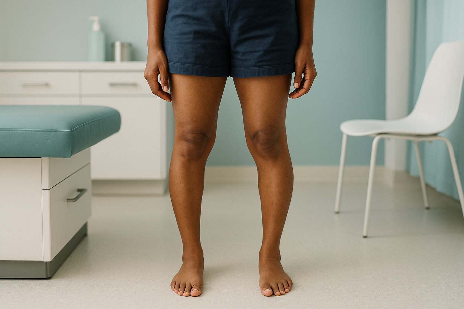
Blount's disease causes a visible inward bowing of one or both shins, usually just below the knee. The bowing is sharper and more angular than the gentle curve you see in a toddler who is still developing. Over time, the affected leg may also develop an inward twist, so the foot points inward. Children and teenagers may limp, complain of knee pain, or simply look "more bowed" than other children their age.
One or both legs bow outward at the knee. The curve deepens over months or years instead of improving.
Pain along the inner knee, sometimes with a feeling that the knee "gives way" while walking or running.
A noticeable limp or waddling walk, especially if only one leg is affected.
The foot on the affected side may turn inward because of tibial torsion (a twist in the shinbone).
How is Blount's disease different from normal bow legs?
Almost all babies are born with some bowing. This is called physiological genu varum. It straightens naturally by around age three or four. In many children, the legs even pass through a mild knock knees phase before settling into their adult alignment.
Blount's disease is different because the bowing does not improve. It gets worse. The growth plate on the inner side of the upper tibia (the proximal tibial physis) stops growing normally, while the outer side keeps growing. This creates a sharper and sharper angle just below the knee. If a child's bow legs are getting worse after age two, or are clearly worse on one side, the child should be assessed.
What causes Blount's disease?
The exact cause is not fully understood. It centres on too much pressure on the growth plate at the top of the tibia. This abnormal load disrupts normal bone development. Several factors raise the risk.
Higher body weight puts more stress on the growth plate. This makes it a strong risk factor for both the infantile and adolescent forms.
Children who walk very early may load the growth plate before it is ready, particularly if they also have higher body weight.
Blount's disease is more common in certain populations and can run in families. This suggests a hereditary part.
Are there different types of Blount's disease?
Yes. Blount's disease is usually grouped by the age at which it appears. The two main forms behave differently and respond to different approaches.
Infantile tibia vara appears before age four. It often affects both legs. If found early, bracing can be effective. If bowing continues beyond age four or is severe, surgery is usually considered. This is the more common form overall.
Adolescent Blount's disease develops after age ten, usually in one leg only. It is strongly linked to higher body weight. Bracing is rarely effective at this stage because much of the bone growth has already happened. Surgery, such as guided growth or an osteotomy (a controlled bone cut to correct the angle), is the usual treatment path.
How is Blount's disease diagnosed with imaging?
Diagnosis relies on a clinical examination plus imaging of the full legs. An x-ray of the upper tibia shows typical changes at the growth plate, including beaking or widening on the inner side. Your specialist will look at the overall leg alignment to measure how severe the bowing is and whether it has changed since the last image.
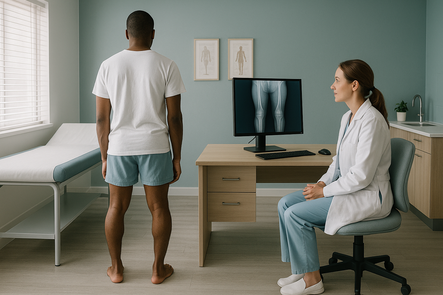
Most importantly, this imaging should be done while the person is standing. When you lie down for a scan, your body weight is taken off the legs and the true bowing angle looks smaller. A full-leg standing x-ray captures the legs from hip to ankle in a single weight-bearing image. This gives the most accurate picture of the deformity.
At ScanAlign, low dose standing imaging, we use EOS technology, which produces standing full-leg images with about 90% less radiation than a standard x-ray, around ten times less. For growing children and teenagers who may need repeat imaging over time, that lower dose matters.
When is MRI the better choice? EOS shows bones, joints and overall alignment clearly. If your specialist needs to look at soft tissues, cartilage, or the growth plate in fine detail, MRI is usually the better tool. In many cases of Blount's disease, both types of imaging are used together.
When do bow legs from Blount's disease need surgery?
Not every case needs an operation. The decision depends on the child's age, how severe the bowing is, and whether it is getting worse. Here is a general guide to the usual steps.
Observation and monitoring
For very young children with mild bowing, the first step is often regular monitoring. This means clinical check-ups and standing imaging to see whether the bowing settles on its own.
Bracing
In infantile Blount's disease (before age four), a knee-ankle-foot brace worn at night may help guide growth. Bracing is less effective if the child is older or if the deformity is severe.
Guided growth surgery
A small plate or staple is placed on the outer growth plate to slow it down for a while. This lets the inner side catch up and straighten the leg. It works best while there is still a lot of growth left.
Osteotomy
For severe cases, or cases found late, the surgeon cuts the tibia and sets it in a straighter position. This is a bigger operation but can correct significant deformity.
ScanAlign is a diagnostic imaging clinic. We do not provide treatment, but our standing full-leg images give your orthopaedic surgeon or specialist the detailed alignment data they need to plan the right approach.
Before your child starts treatment, or changes the one they are on, it is worth getting a detailed diagnosis. Knowing whether the cause sits in the pelvis, the legs or the spine changes what will actually help.
Worried that the bowing is getting worse? A standing full-leg image gives your specialist the clearest picture of the deformity.
Book a children's leg alignment scanFrequently asked questions about Blount's disease
Most children's bow legs straighten naturally by age three to four. If bowing is still there or getting worse after this age, it is worth having the legs assessed by a specialist.
Normal bowing is the same on both sides and slowly improves. Blount's disease usually causes a sharper, more angular bow just below the knee. It may affect one leg more than the other, and it gets worse over time instead of better.
No. Rickets is caused by a lack of vitamin D or calcium, which softens all the bones. Blount's disease is a growth plate problem in one place, at the top of the tibia. They look different on imaging, though both can cause bowing.
The infantile form often affects both legs. The adolescent form more commonly affects just one. Even when both legs are involved, the severity can differ between them.
Blount's disease is diagnosed during growth. However, adults who had untreated or partly treated Blount's disease as children may still have some bowing and related joint problems that benefit from an alignment assessment.
When you lie down, gravity no longer acts on the bones. A standing image shows the real angle of bowing under your full body weight. This gives the surgeon the most accurate measurement to plan treatment.
EOS uses about 90% less radiation than a standard x-ray, around ten times less. This is particularly helpful when a young person needs repeat imaging to monitor the deformity over time.
No. You can book directly yourself. We offer a free video consultation first so we can understand the concern and confirm that a standing full-leg image is the right next step.
The price for patients travelling from abroad is confirmed during the free video consultation.
It can come back, particularly in the infantile form or if the child still has a lot of growth left. Regular monitoring with standing imaging helps pick up any return of bowing early.
Worried that your child's leg bowing is getting worse?
A free video consultation lets us understand the concern and advise whether a standing full-leg image is the right next step before you plan a trip to London.
Book a free video consultationSources
- Sabharwal S. Blount disease. Journal of Bone and Joint Surgery (American). 2009;91(7):1758-1776.
- NHS. Bow legs. NHS Health A to Z. Available at: nhs.uk.
- Langenskiöld A. Tibia vara (osteochondrosis deformans tibiae): a survey of 23 cases. Acta Chirurgica Scandinavica. 1952;103(1):1-22.
Written by Abbas Dhami (Specialist Diagnostic Radiographer)
This page is general information and not a diagnosis.
ScanAlign is the trading name of CoreMed Solutions Ltd and operates under The Harley Street Hospital's CQC licence.