
EOS or DEXA: Which Scan Answers Which Question?
A DEXA scan measures bone mineral density. It tells you how strong your bones are and whether you are at risk of osteoporosis. An EOS scan shows the shape, position and alignment of your bones while you stand. The two scans answer completely different questions. So the right choice depends on what you need to know.
Key points
- DEXA measures bone density (strength). EOS maps bone alignment (shape and position).
- A DEXA scan cannot show whether your skeleton is aligned properly. An EOS scan cannot measure bone density.
- Some people benefit from both scans, especially after a fracture, or when ongoing pain is being looked into alongside known low bone density.
- EOS uses about 90% less radiation than a standard X-ray, around ten times less.
- Neither scan replaces MRI when soft tissue, discs or nerves need to be assessed.
What does a DEXA scan show compared with an EOS scan?
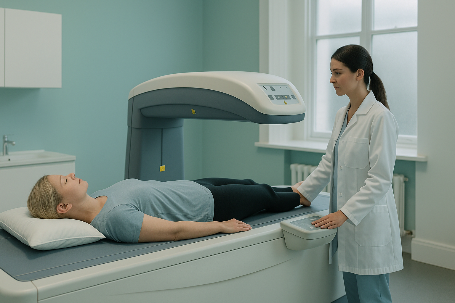
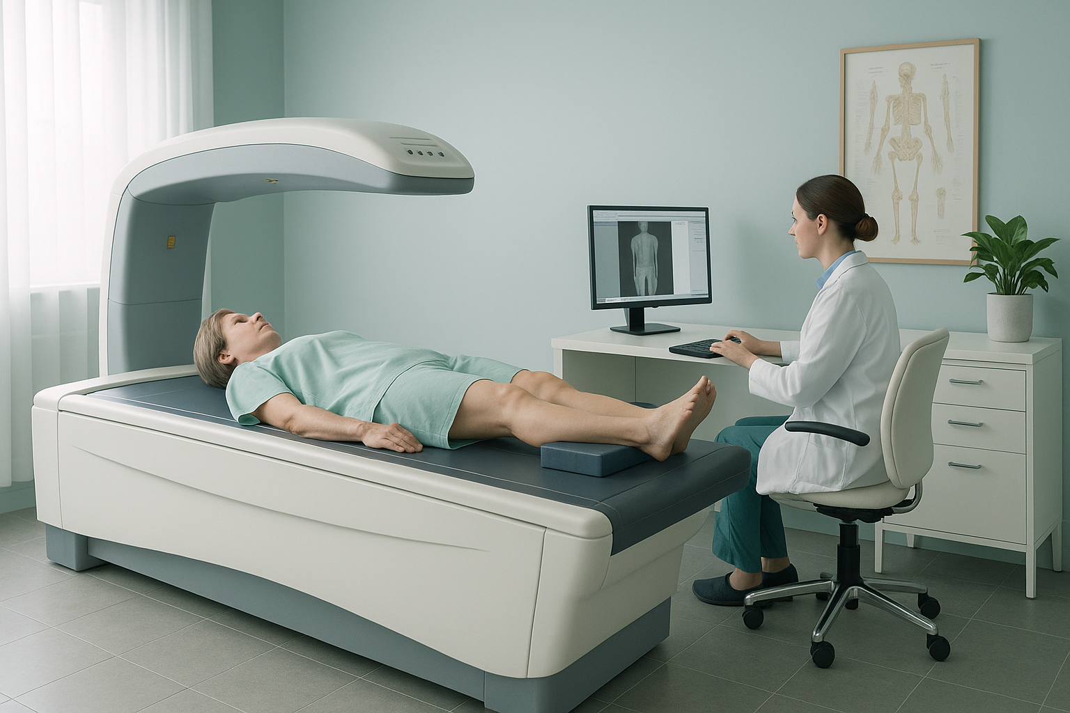
A DEXA scan (dual-energy X-ray absorptiometry) measures how much mineral is packed into a section of bone. This is usually the hip and lower spine. The result is a T-score. It tells your doctor whether your bone density is normal, low (osteopenia) or very low (osteoporosis). It is the standard test for fracture risk.
An EOS scan does something entirely different. It takes a full-length image of your skeleton while you stand, carrying your own weight. This shows how your spine curves, whether your pelvis is level, and how your hips, knees and ankles line up under real-life load. You can read more about EOS compared with X-ray, CT and MRI.
How do DEXA and EOS scans compare side by side?
The table below sets out the main differences. The key point is that these scans measure different things. One does not replace the other.
| Feature | DEXA scan | EOS scan |
|---|---|---|
| What it measures | Bone mineral density (strength) | Bone shape, position and alignment |
| Position during the scan | Lying down | Standing up, weight-bearing |
| Main clinical use | Diagnosing osteoporosis and assessing fracture risk | Mapping whole-body skeletal alignment under load |
| Body areas covered | Usually hip and lower spine | Full length, head to feet |
| Radiation | Very low | About 90% less than a standard X-ray, around ten times less |
| Shows soft tissue or discs | No | No |
| Shows alignment under gravity | No | Yes |
When should you choose a DEXA scan and when should you choose EOS?
The choice depends entirely on the question that your clinician (or you) needs answered.
- You need to check for osteoporosis or osteopenia
- You are tracking bone density over time, for example during treatment
- You want a fracture-risk assessment based on a T-score
- A condition such as osteomalacia has raised concerns about bone strength
- You have ongoing back, hip or knee pain that standard imaging has not explained
- You need a full picture of how your skeleton lines up while standing
- A surgeon or specialist needs weight-bearing measurements before planning treatment
- You want to understand whether misalignment in one area is causing pain elsewhere
A note on MRI. EOS shows bones, joints and alignment. For soft tissue, discs or nerves, MRI is usually the better tool. If your symptoms include nerve pain, numbness or disc problems, your clinician may recommend MRI instead of, or alongside, an EOS scan.
Do some people need both a bone density scan and an alignment scan?
Yes. Bone density and bone alignment are separate pieces of the puzzle. Knowing that your bones are weaker than expected (DEXA) is important. But it does not tell you whether those weaker bones are also carrying load unevenly because your spine or pelvis is out of line (EOS).
After a vertebral fracture, for example, a DEXA scan confirms the bone density problem. A standing EOS scan then shows how that fracture has changed your overall alignment, and whether your body is compensating in ways that could lead to further problems. The body is a connected chain from feet to head. A change in one place often shifts the load somewhere else.
If your clinician is looking into both bone strength and skeletal shape, the two scans complement each other rather than overlap.
Not sure which type of imaging you need? Our radiographer can help you work it out.
Book a free video consultationHow do you book a standing alignment scan at ScanAlign?
You do not need a referral from your doctor. You can book a standing alignment scan yourself in four simple steps, before you plan your trip to London.
Speak with our specialist radiographer by video from home. He will listen to your symptoms, review any previous imaging and confirm whether an EOS scan is the right next step.
The scan itself takes only a few minutes. You stand naturally inside the EOS system while it takes full-length images from head to feet, using about 90% less radiation than a standard X-ray, around ten times less.
A specialist radiologist analyses the images and writes a detailed alignment report. It covers your spine, pelvis, hips, knees and ankles.
You receive the report and images. You can share them with any clinician involved in your care, including your clinicians back home. ScanAlign is a diagnostic imaging clinic and does not provide treatment, but the findings give your treating team the information they need.
Before you start a treatment, or change the one you are on, it is worth getting a detailed diagnosis first. Knowing whether the cause sits in your pelvis, your legs or your spine changes what will actually help.
Frequently asked questions
A DEXA scan measures bone mineral density and gives a T-score, which is used to diagnose osteoporosis. An EOS scan does not measure density. It shows the shape and alignment of your bones while they carry your weight.
No. They answer different questions. If you need to know how strong your bones are, you need a DEXA scan. If you need to know how your skeleton lines up under load, you need an EOS scan.
Yes. An EOS scan uses about 90% less radiation than a standard X-ray, around ten times less. This makes it one of the lowest-dose skeletal imaging options available.
No. ScanAlign accepts adults who refer themselves. You can book directly through a free video consultation with our specialist radiographer.
ScanAlign is a private self-pay clinic. For patients travelling from abroad, the price is confirmed during the free video consultation. You can also visit our FAQs and cost page.
In the UK, DEXA scans are widely available on the NHS (usually through a GP referral) and at many private clinics. ScanAlign does not offer DEXA scans. We provide ScanAlign standing EOS imaging to assess skeletal alignment.
A DEXA scan can find low bone density, which may contribute to fractures that cause pain. However, it cannot show alignment problems, disc problems or soft-tissue causes of back pain. A standing alignment scan or MRI may be more suitable, depending on your symptoms.
Bone density means how much mineral is packed into the bone, which decides how strong it is. Bone shape means the structure and position of the bones in your skeleton. You can have normal density but poor alignment, or low density with good alignment. They are separate measurements.
When you lie down, gravity is no longer compressing your spine and joints. Your muscles relax and your skeleton shifts into a different position. A standing scan captures the alignment your body actually uses through the day. This can reveal problems that lying-down imaging misses.
You speak with Abbas Dhami, our specialist diagnostic radiographer. He will ask about your symptoms and any previous imaging. He will explain what an EOS scan can and cannot show, and help you decide whether a standing alignment scan is the right next step for you.
Not sure which scan you need?
Our specialist radiographer will talk you through your options in a free video call, with no obligation.
Book a free video consultationWritten by Abbas Dhami (Specialist Diagnostic Radiographer)
Sources
- NHS. "DEXA (DXA) scan." nhs.uk. Accessed 2025.
- Deschênes S, et al. "Diagnostic imaging of spinal deformities: reducing patients' radiation dose with a new slot-scanning X-ray imager." Spine, 2010;35(9):989-994.
- International Society for Clinical Densitometry (ISCD). "Official Positions: Adult." iscd.org. Accessed 2025.
This page is general information and not a diagnosis.
ScanAlign is the trading name of CoreMed Solutions Ltd and operates under The Harley Street Hospital's CQC licence.