
Disc Bulge or Disc Herniation: What Is the Difference?
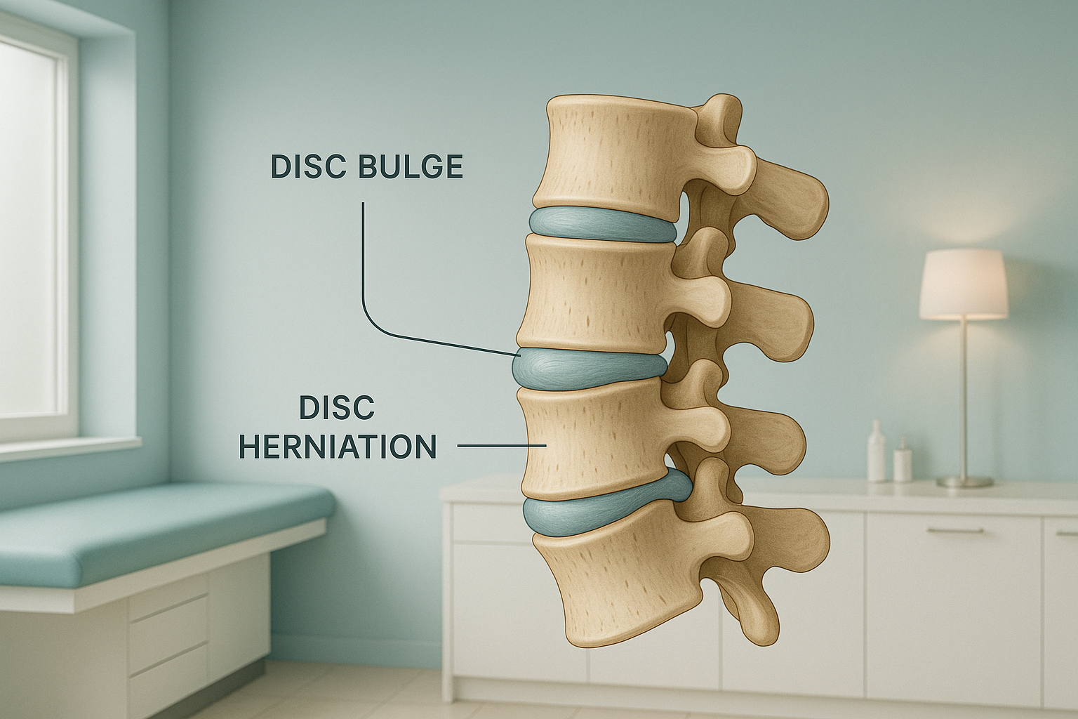
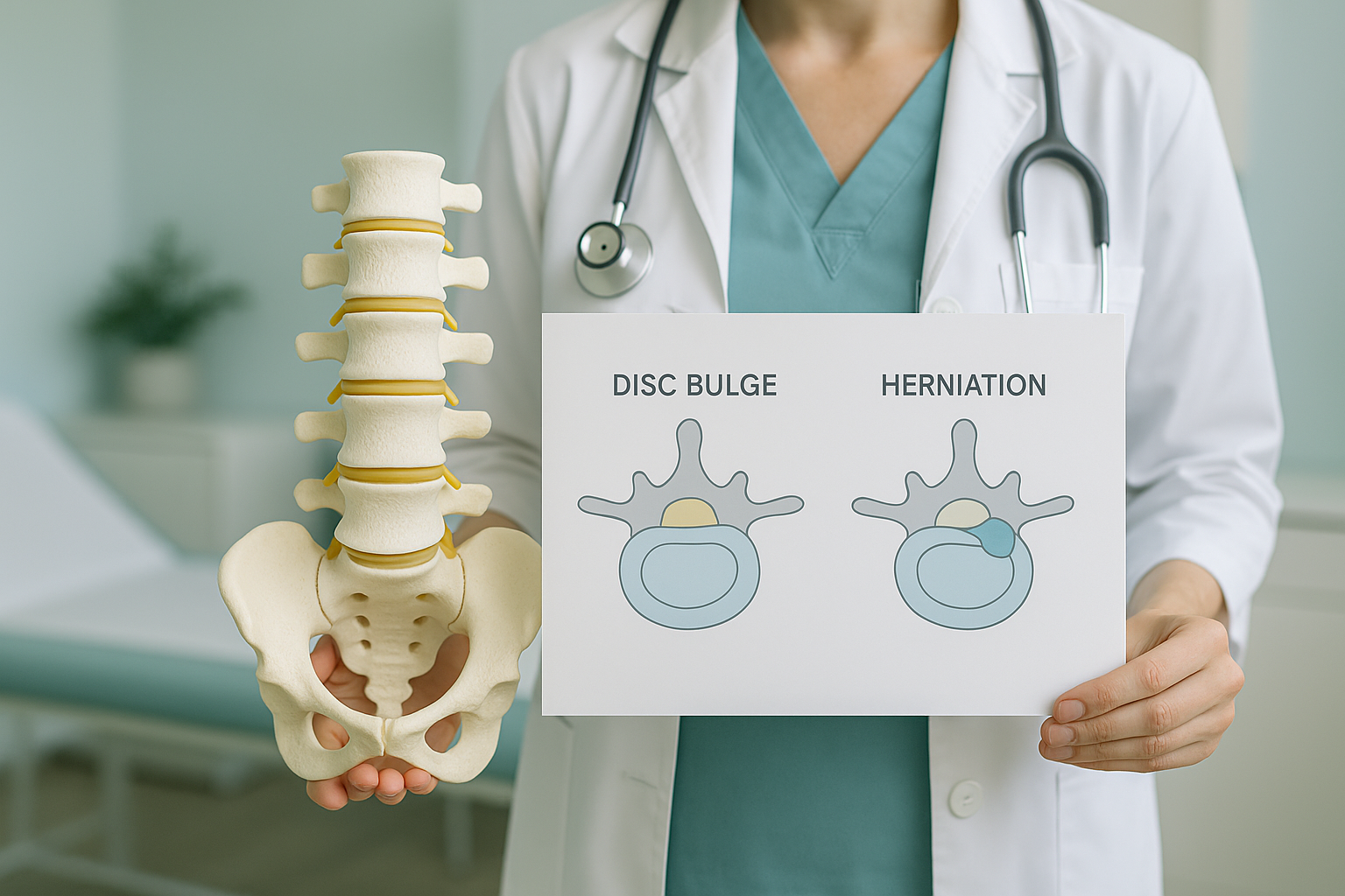
A disc bulge means the outer wall of a spinal disc has spread outward over a wide area, like a tyre low on air. A disc herniation means the soft centre has pushed out through a tear in that outer wall. A bulge is usually wider and flatter. A herniation is more focused in one spot and more likely to press on a nearby nerve. Still, many bulges and herniations cause no pain.
Key takeaways
- In a bulge, the whole disc wall bows outward. In a herniation, the inner material breaks through in one spot.
- Both can show up on scans in people who have no symptoms at all.
- When symptoms do occur, a herniation is more likely to cause a pinched nerve, because it sticks out further.
- Lying-down scans can make a disc look less loaded than it really is. Standing imaging shows the spine under real weight.
- Most disc problems get better over time without surgery.
What does a spinal disc actually look like inside?
Each spinal disc is a tough, flexible pad that sits between two vertebrae. It has two parts. The outer ring, called the annulus fibrosus, is made of layers of fibrous tissue, a bit like the layers of a car tyre. The inner core, the nucleus pulposus, is a gel-like substance. It absorbs shock when you move, bend or carry weight.
If the outer ring stays whole but starts to sag outward, that is a bulge. If the outer ring tears and the gel pushes through, that is a herniation. The difference matters. A herniation can release inflammatory chemicals near nerve tissue, even if the bulge itself is small.
What are the different types of disc problem?
Disc problems sit on a scale. A bulge is the mildest form. A herniation can be graded further, based on how far the inner material moves. You may see these words on a scan report.
The disc wall bows outward over a wide area, often more than half the way around the disc. The outer ring is still whole.
A type of herniation where the base of the bulge is wider than the part that sticks out. The outer ring may be weakened, but it still partly holds the material in.
The inner gel has broken fully through the outer ring. The part that sticks out is narrower at its neck than at its tip.
A piece of disc material has broken off completely and now lies free in the spinal canal. This is the most advanced stage.
Many reports also mention loss of disc height. This is a sign of degenerative disc disease, the slow wear that makes discs more likely to bulge or herniate.
Does a bulging or herniated disc always cause pain?
No. Research shows again and again that many adults have disc bulges, or even herniations, on MRI but feel no pain at all. The finding on its own does not mean you need treatment. Symptoms tend to start when the disc material presses on or irritates a nearby nerve root. This is often called nerve root compression.
Common symptoms when a disc does cause trouble
- Back or neck pain in one area that gets worse with sitting or bending forward.
- Pain, tingling or numbness spreading into the arm or leg, depending on which level is affected.
- Muscle weakness in the affected arm or leg.
- Stiffness that eases with gentle movement but comes back after staying in one position for a long time.
When to seek urgent help
If you suddenly lose control of your bladder or bowels, have numbness around the groin, or have weakness in both legs that is getting worse quickly, get urgent medical help. Contact your doctor or go to the nearest emergency department. These signs may point to a rare but serious condition called cauda equina syndrome.
What causes a disc to bulge or herniate?
Discs are under load all the time. Over years of bending, lifting and sitting, the outer ring can weaken. Several things speed this up.
Repeated bending, heavy lifting or long hours of sitting add strain over time.
Discs naturally lose water and flexibility as you get older.
A fall, an awkward twist or a heavy lift can tear the outer ring suddenly.
Poor posture or a tilt in the pelvis can put more load on one side of a disc than the other.
Often there is no single big event. The disc weakens slowly, and then a small movement sets off the final bulge or tear.
How does standing imaging help with disc problems?
Standing, weight-bearing imaging shows your spine under the same load it carries all day. When you lie down for a usual scan, gravity is taken away. The muscles relax and the vertebrae shift. A disc that bulges under load may then look less severe, or even normal, on a lying-down image.
What standing shows. When you are upright, gravity presses down on the discs and loads the joints. Any bulge, narrowing or shift can be seen as it really is when you move and work. The alignment of the whole spine, from the skull to the pelvis, is captured in one image. Your clinician can then see whether a tilt or curve somewhere else is adding load to the affected disc.
What lying down can miss. Lying flat takes the weight off the spine. Discs decompress slightly, vertebrae may drift back to a more neutral position, and a misalignment that causes problems during the day can disappear. The scan may report "mild" findings that are actually more significant when you stand.
An EOS scan at ScanAlign captures a full-length standing image of your spine with about 90% less radiation than a standard X-ray, around ten times less. It does not replace MRI, but it adds the weight-bearing view that MRI cannot give.
A note on MRI. EOS shows bones, joints and alignment under load. For soft tissue detail, such as the disc material itself, the spinal cord or the nerve roots, MRI is usually the better tool. The two work well together. If your clinician needs to see both structure and alignment, putting MRI findings together with a standing EOS image gives the fullest picture.
Wondering whether a standing spine image could help explain your symptoms? You can book a standing spine assessment with a free video consultation first, from home, so we can check it is the right step for you before you travel to London.
Book a free video consultationCan a bulging or herniated disc get better on its own?
Yes, in many cases. Research shows that herniated disc material can shrink over time through a process called natural resorption, where the body slowly absorbs the tissue that sticks out. Many people find their symptoms ease a lot within weeks to months with conservative care, such as physiotherapy, gentle movement and pain management guided by their doctor or specialist.
Accurate imaging helps your clinician choose the best path. If a structural misalignment is adding extra load to the affected disc, finding and addressing it may be an important part of the plan.
So before you start treatment, or change the one you are on, it is worth getting a detailed diagnosis. Knowing whether the cause sits in the pelvis, the legs or the spine changes what will actually help.
Frequently asked questions
Generally, yes. A herniation means the inner gel has broken through the outer wall, so it is more likely to press on a nerve. But how serious it is depends on where it is and what symptoms it causes, not just the label. Many herniations cause no pain.
It can. If the outer ring keeps weakening, the inner gel may in time push through. This does not always happen, and many bulges stay stable for years.
A disc protrusion is a type of herniation where the base of the displaced material is wider than the part that reaches outward. It is a middle stage, between a simple bulge and a full extrusion.
Most herniated discs get better without surgery. Your doctor or specialist will consider surgery only if symptoms are severe, getting worse, or not responding to other treatments over several months.
Disc bulges and herniations are very common findings in people without symptoms, especially over the age of 40. The scan finding alone does not mean you need treatment. Symptoms matter more than the image.
In an extrusion, the inner gel has broken through the outer ring but is still attached to the disc. In a sequestration, a piece has broken off completely and lies loose in the spinal canal.
EOS shows bones and alignment, not soft tissue detail. It will not show the disc material itself. However, it can show loss of disc height, shifts in the vertebrae, and overall alignment problems that add to disc overload. MRI is the standard tool for seeing the disc directly.
Yes. EOS uses about 90% less radiation than a standard X-ray, around ten times less. It is a very low-dose imaging system.
No. You can refer yourself. The process starts with a free video consultation to make sure the scan is right for you.
The price for patients travelling from abroad is confirmed during the free video consultation, and our FAQs page answers other common questions.
Understand how your spine is loaded
A standing whole-body image shows your clinician the full picture. Start with a free video consultation from home to see if it is the right step for you.
Book a free video consultationSources
- Brinjikji W, Luetmer PH, Comstock B, et al. Systematic literature review of imaging features of spinal degeneration in asymptomatic populations. American Journal of Neuroradiology. 2015;36(4):811-816.
- Fardon DF, Williams AL, Dohring EJ, et al. Lumbar disc nomenclature: version 2.0. The Spine Journal. 2014;14(11):2525-2545.
- National Institute for Health and Care Excellence (NICE). Low back pain and sciatica in over 16s: assessment and management. NICE guideline NG59. 2016 (updated 2020).
Written by Abbas Dhami (Specialist Diagnostic Radiographer)
This page is general information and not a diagnosis.
ScanAlign is the trading name of CoreMed Solutions Ltd and operates under The Harley Street Hospital's CQC licence.