
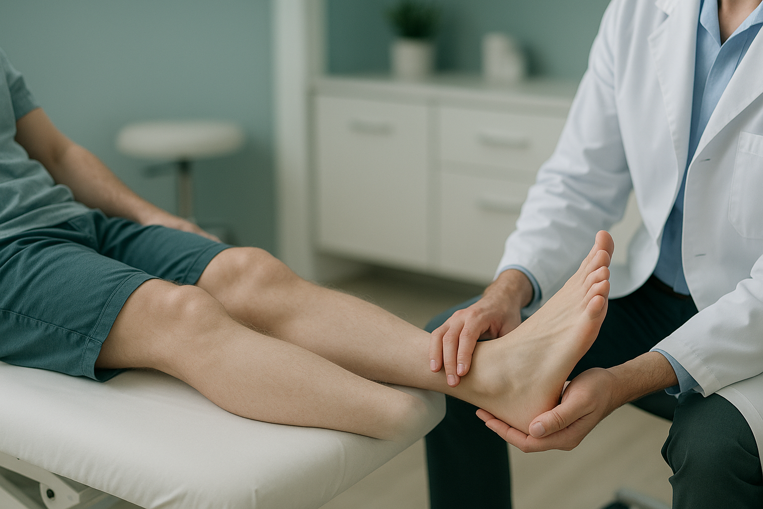
What Causes Foot Drop, and How Urgent Is It?
Foot drop means it is hard to lift the front of your foot. The usual cause is pressure on the common peroneal nerve at the knee, or nerve root compression in the lower back, most often at the L5 level. New or worsening foot drop needs a prompt medical check, because early diagnosis improves recovery.
Key points
- Foot drop means you cannot lift your toes and the front of your foot properly when you walk.
- The most common causes are a peroneal nerve injury at the knee or a compressed nerve root in the lower back.
- New foot drop with numbness, weakness, or bladder or bowel changes is a medical emergency.
- Standing whole-body imaging can show spinal alignment problems that lying-down scans may miss.
- An early check gives you the best chance of recovery.
How urgent is foot drop?
It depends on how fast it appeared and whether you have other symptoms. Foot drop that comes on suddenly may point to a serious spinal problem, such as cauda equina syndrome. This is especially true if it comes with back pain, leg weakness, or changes in bladder or bowel control. This needs emergency care.
Get urgent medical help (your doctor or the emergency department of your nearest hospital) if you have new foot drop together with any of the following:
- Numbness around the groin or inner thighs (saddle area)
- Loss of bladder or bowel control
- Leg weakness that is getting worse quickly
- Sudden severe back pain
Do not wait for a private scan. These symptoms need to be checked straight away.
If your foot drop has come on slowly and you have none of these warning signs, it still needs proper investigation. The longer a nerve is compressed, the harder recovery becomes. Speak to your doctor or a specialist soon.
How do you know if you have foot drop?
You may notice that you cannot lift the front of your foot properly. Your toes may drag or catch on the ground when you walk. Many people change the way they walk without realising it.
Your toes catch on kerbs, steps or uneven ground more than they used to.
You lift your knee higher than normal so your foot does not drag. This is sometimes called a steppage gait.
Your foot slaps down on the floor when you step, because you cannot control the landing.
Less feeling over the top of the foot or the outer shin, which suggests a nerve is involved.
What causes foot drop?
Foot drop is a symptom, not a condition in itself. It happens when the nerve signal that tells your foot to lift is interrupted somewhere along its path, from the lower back down to the knee. The two most common trouble spots are the common peroneal nerve on the outside of the knee and the L5 nerve root in the lumbar spine.
The nerve wraps around the head of the fibula, just below the knee. Here it can be hurt by pressure from tight casts, crossing your legs, or a direct injury.
A bulging or herniated disc in the lower back can compress the L5 nerve root. This causes L5 radiculopathy, with weakness in the muscles that lift the foot.
Narrowing of the spinal canal can slowly squeeze nerve roots. This is more common with age and can cause lumbar radiculopathy.
Less common causes include stroke, motor neurone disease, nerve damage from diabetes, and complications of hip or knee surgery.
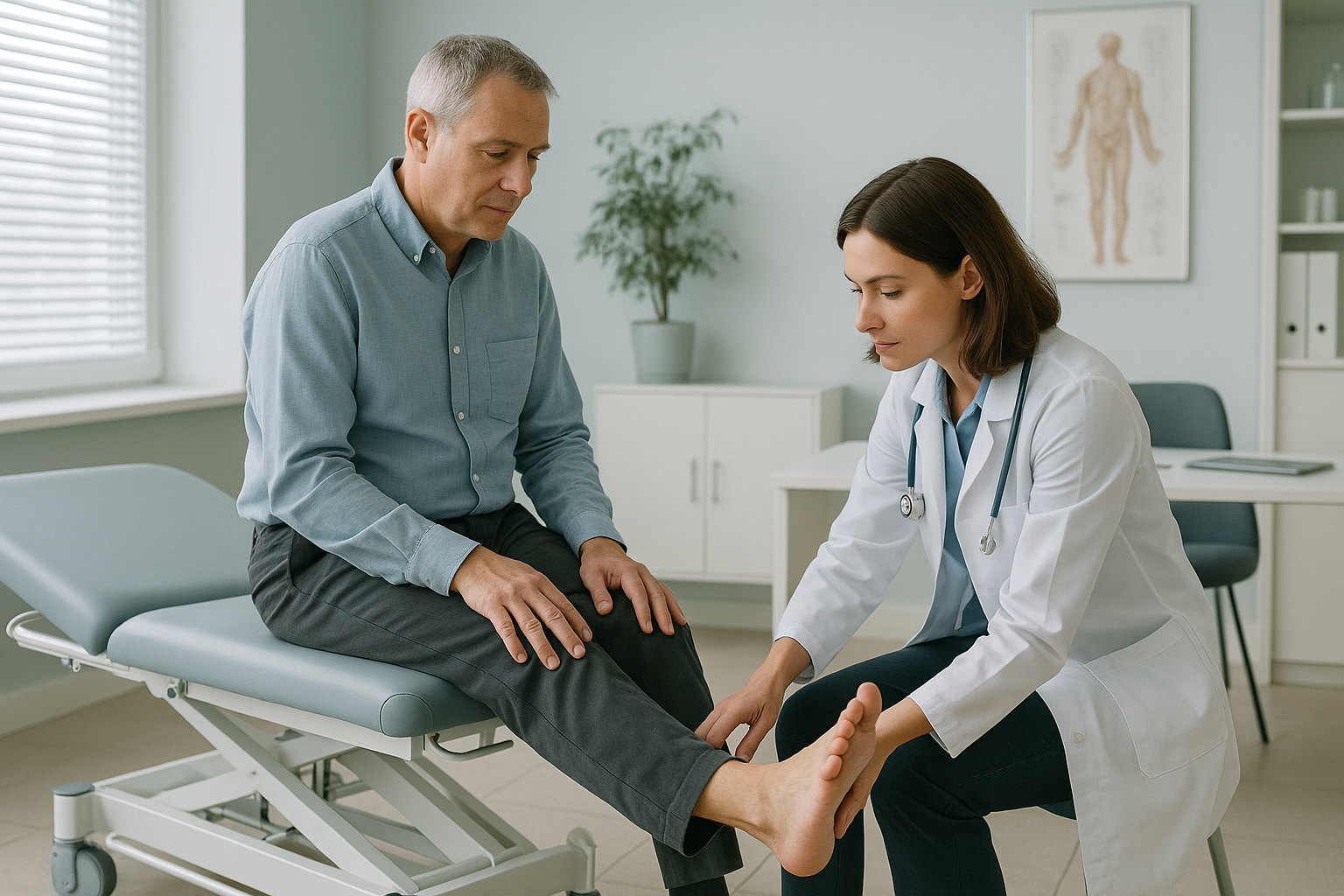
The nerve travels a long way from the spine to the foot. So the cause of foot drop is not always where you feel the symptoms. A problem in your lower back can cause weakness at your ankle. This is why a full assessment matters.
How does standing imaging help with foot drop?
Standing imaging shows your spine and legs under the same load they carry all day. When you lie down for a standard X-ray, CT or MRI, your muscles relax and gravity is taken away. Alignment problems that only show up when you bear weight can be hidden.
Your body works as a linked chain from your feet to your head. If your pelvis tilts or your spine shifts to one side, the nerve roots in your lower back can come under pressure that a lying-down image would not show. ScanAlign standing EOS imaging takes a full-length image of your skeleton while you stand naturally, using about 90% less radiation than a standard X-ray, around ten times less.
For someone whose foot drop has not been fully explained by other tests, a standing whole-body assessment can show spinal or pelvic alignment shifts that may be adding to the nerve compression.
When MRI is the better tool: EOS imaging shows bones, joints and alignment. For soft tissue detail, such as a herniated disc pressing on a nerve, MRI is usually the better test. In many cases, standing alignment imaging and MRI together give the fullest picture.
If your foot drop has been investigated but the cause is still unclear, a standing whole-body assessment may show alignment factors that other imaging missed.
Book a free video consultationWhat should you do if you think you have foot drop?
The right first step depends on how quickly your symptoms appeared.
- See your doctor urgently or go to the emergency department
- Mention any back pain, numbness or bladder changes
- You may need an emergency MRI and a specialist review
- You may already have had an MRI, nerve conduction studies or physiotherapy
- Standing alignment imaging can check whether spinal or pelvic alignment is playing a part
- A free video consultation can help you decide if this is the right next step before you plan a trip to London
Recovery from foot drop varies. It depends on the cause, how long the nerve has been compressed, and how soon treatment starts. Some people recover fully. Others need ongoing support, such as an ankle-foot orthosis to help with walking. ScanAlign does not provide treatment. It provides the diagnostic imaging that helps your treating clinician plan the right approach.
Before you start a treatment, or change the one you are on, it is worth getting a detailed diagnosis. Knowing whether the cause sits in your pelvis, your legs or your spine changes what will actually help.
If your symptoms include nerve root compression in the lower back, understanding your whole-body alignment can be an important part of the diagnosis. You can book an assessment to talk through your situation with a specialist radiographer first.
Frequently asked questions about foot drop
It can, especially if the cause is short-term pressure on the peroneal nerve, for example from a cast or a habit of crossing your legs. If a compressed nerve root in the spine is the cause, recovery depends on relieving that pressure. The sooner the cause is found, the better the outlook.
It can be. Many cases are caused by nerve compression that can be treated. But foot drop can occasionally be a sign of stroke, motor neurone disease or cauda equina syndrome. New foot drop should always be checked by a doctor.
The peroneal nerve can be compressed at the knee. This causes weakness limited to the foot and outer shin. An L5 nerve root problem starts in the lower back. It can cause wider symptoms, including back pain, hip weakness and numbness down the leg, as well as foot drop.
No. ScanAlign accepts self-referrals. You start with a free video consultation, where a specialist radiographer reviews your situation before any scan is booked.
An EOS scan uses about 90% less radiation than a standard X-ray, around ten times less. This makes it especially suitable when full-body images are needed.
When you lie down, gravity is taken away and your muscles relax. Spinal alignment shifts that happen under normal standing load, such as a pelvic tilt or a lean to one side, may flatten out. If these shifts are adding to nerve compression, a lying-down scan may not show them.
EOS imaging shows bone and joint alignment under load. It can show spinal or pelvic alignment problems that add to nerve compression. But to see soft tissue, such as a herniated disc, an MRI is usually needed as well. The two tests work well together.
ScanAlign is a private, self-pay clinic. The price for patients travelling from abroad is confirmed during the free video consultation.
Common tests include an MRI of the lumbar spine, nerve conduction studies (which test electrical signals in the nerve), and standard X-rays. Standing whole-body imaging adds alignment information that other tests do not capture.
A specialist radiographer will listen to your history, review any previous imaging you have, and explain whether a standing alignment assessment is likely to add useful information in your case. There is no obligation to go ahead.
Still have questions about foot drop?
A free video consultation with a specialist radiographer can help you understand whether standing alignment imaging is the right next step, before you decide to travel to London.
Book a free video consultationWritten by Abbas Dhami (Specialist Diagnostic Radiographer)
Sources
- NHS. "Foot drop." nhs.uk. Accessed 2024.
- Stewart JD. "Foot drop: where, why and what to do?" Practical Neurology. 2008;8(3):158-169.
- American Academy of Orthopaedic Surgeons. "Foot Drop." OrthoInfo. Accessed 2024.
This page is general information and not a diagnosis.
ScanAlign is the trading name of CoreMed Solutions Ltd and operates under The Harley Street Hospital's CQC licence.