
How Do You Read and Understand Your Radiology Report?
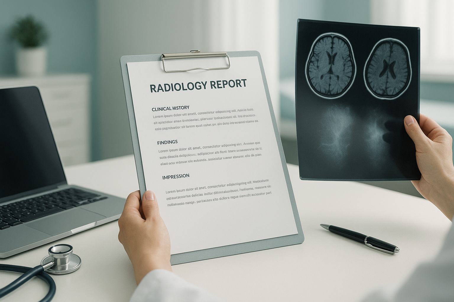
A radiology report is a written summary by a radiologist. It describes what your scan shows. It usually has four parts: clinical details, findings, a comparison with any previous imaging, and an impression (the main conclusion). Terms like "unremarkable" mean normal, and "degenerative change" means age-related wear. The doctor who sent you for the scan uses the report to guide your next steps.
Key takeaways
- Every radiology report follows a standard structure, from clinical history to impression.
- Most confusing terms have simple meanings in plain English.
- "Clinical correlation recommended" is not a red flag. It means your doctor should match the findings to your symptoms.
- Standing imaging can reveal alignment problems that lying-down scans miss.
- Always discuss your report with the clinician who asked for the scan.
You have had a scan. Now you are looking at a report full of unfamiliar words. It can feel confusing, even worrying, when you do not know what the terms mean. This page goes through a standard radiology report section by section. It explains the most common phrases in everyday language, so you can go into your next appointment feeling informed.
What are the main sections of a radiology report?
Most radiology reports follow the same four-part layout, whether the scan was an X-ray, CT, MRI or EOS image. Knowing the layout helps you find the information that matters most.
1. Clinical details
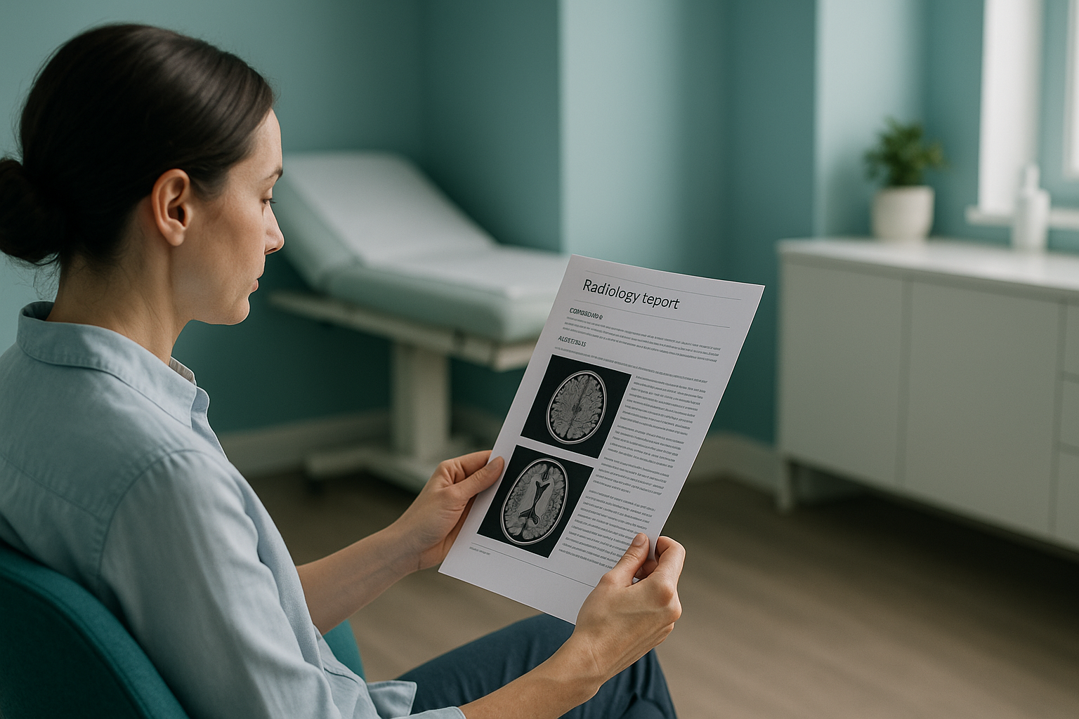
This short paragraph explains why the scan was requested. It usually includes your symptoms, the referring clinician's question (for example, "query fracture" or "low back pain") and any relevant history. If this section is vague, the radiologist has less context. This can affect how they interpret the images.
2. Findings
This is the longest section. The radiologist describes what they see, area by area. They may note the condition of bones, joints, discs or soft tissue. This is where you will meet most of the technical language, which we explain below.
3. Comparison with prior imaging
If you have had a previous scan, the radiologist may compare the two. They will note whether anything has changed, stayed the same or improved. If there is no earlier scan, this section will say "no prior imaging available for comparison."
4. Impression
This is the summary. It is usually the most useful part for you and your clinician. It brings the key findings together into a short conclusion. Sometimes it suggests what to do next, such as "clinical correlation recommended" or "further imaging advised."
What do common radiology report terms mean in plain English?
Radiologists use precise, standard language so that any doctor reading the report knows exactly what was seen. Here is a glossary of the phrases that cause the most confusion.
| Report term | What it actually means |
|---|---|
| Unremarkable | Normal. Nothing unexpected or abnormal was seen in that area. |
| Degenerative change | Wear and tear related to age. Very common, especially in the spine. Often seen in people with no symptoms at all. Read more about degenerative disc disease. |
| Incidental finding | Something noticed that was not the reason for the scan. It may or may not be important. Your clinician will decide if it needs follow-up. |
| Clinical correlation recommended | The radiologist is saying the images alone do not tell the whole story. Your doctor needs to match the finding with your symptoms and examination. |
| Osteophyte | A small bony growth (bone spur) at the edge of a joint. A sign of wear, not necessarily a cause of pain. |
| Disc bulge / protrusion | A disc between the vertebrae is pushing outward slightly. Very common on MRI, and many people with disc bulges have no symptoms. |
| Spondylolisthesis | One vertebra has slipped forward or backward compared with the one below it. Learn more about spondylolisthesis. |
| Effusion | Extra fluid inside a joint. It can indicate inflammation or injury. |
| No acute abnormality | Nothing new or urgent was found. There may still be long-standing changes noted elsewhere in the report. |
| Maintained alignment | The bones are in their expected position. Nothing is shifted or displaced. |
If your report mentions a term you do not recognise, write it down. Ask your referring clinician to explain it at your next appointment. The radiologist writes for a medical audience, so some explaining is to be expected.
What do mild, moderate and severe mean on a scan report?
These grading words describe how large or advanced a finding is. But they are more subjective than you might think. There is no universal cut-off that separates mild from moderate. Different radiologists may grade the same image slightly differently.
A small or early change. Often age-related and not necessarily linked to your symptoms.
A more noticeable change. May or may not cause symptoms, depending on the area and your overall alignment.
A significant change that is more likely to be relevant. Your clinician will decide the best course of action.
The key point is this: the grade on a report does not always match how you feel. Some people with "severe" degenerative change have no pain. Others with "mild" findings are very uncomfortable. That is why the impression often says "clinical correlation recommended." The scan is one piece of the puzzle, not the whole picture.
A note on MRI vs bone imaging. EOS and X-ray reports focus on bones, joints and alignment. MRI is usually the better tool for soft tissue, discs and nerves. If your report mentions a disc or nerve issue, that was likely found on MRI. Both types of imaging have a role, and they often complement each other.
Why does it matter whether your scan was taken standing or lying down?
It matters because gravity changes how your body is aligned. When you lie down for a standard X-ray, CT or MRI, your muscles relax. Your spine, pelvis and joints settle into a position you never actually use in daily life. Small shifts, tilts and imbalances that only appear under your body weight can disappear on a lying-down scan.
Your body works as a connected chain from your feet to your head. A tilt at the pelvis can make the spine compensate. That compensation can show up as pain in the lower back, the hip, or even the knee. A lying-down scan may show each area as "unremarkable" on its own, yet miss the overall alignment problem that is driving your symptoms.
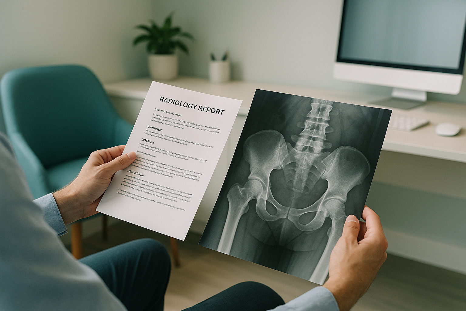
Standing whole-body imaging, such as an EOS scan, captures your skeleton from head to feet while you stand naturally. The report from this type of scan includes alignment measurements across your whole frame, not just one area on its own. We have a dedicated page on how EOS scan results are reported.
Thinking about travelling to London for a standing whole-body assessment? With ScanAlign standing EOS imaging at 19 Harley Street, London, you get a detailed report that maps your alignment from head to feet under natural weight-bearing conditions.
Book a free video consultationWhat should you do after receiving your scan results?
The most important step is to discuss the report with the clinician who asked for the scan. They can explain how the findings relate to your symptoms and what, if anything, needs to happen next.
Read the impression first
Go straight to the bottom of the report. The impression gives you the main conclusion without the technical detail.
Note any terms you do not understand
Write them down. Use the glossary above or bring them to your next appointment.
Ask your clinician the right questions
"Is this finding likely to be causing my symptoms?" and "Do I need any further imaging?" are good starting points.
Consider whether alignment has been assessed
If your report was from a lying-down scan and your symptoms persist, standing whole-body imaging may reveal what was missed. See what an EOS scan report shows for more detail.
Before you start a treatment, or change the one you are on, it is worth getting a detailed diagnosis. Knowing whether the cause sits in your pelvis, your legs or your spine changes what will actually help.
A word of caution. If you have sudden numbness, weakness, or any loss of bladder or bowel control along with your back or joint pain, do not wait for a scan appointment. Contact your doctor, call your local emergency number, or go to the nearest emergency department immediately. These are signs that need urgent medical attention.
Frequently asked questions about radiology reports
It means normal. The radiologist did not see anything unexpected or abnormal in that area. It is a positive finding, even though the word sounds dismissive.
It means the scan finding needs to be matched with your symptoms and physical examination. The radiologist is pointing out that the images alone are not enough to reach a final diagnosis. Your referring clinician will make that decision.
Not necessarily. Degenerative change is extremely common and is essentially wear and tear. Many people have it on their scans with no symptoms at all. Whether it is relevant depends on where it is and what symptoms you have. Your clinician can help you understand whether it is connected to your problem.
Yes. Under UK law you have the right to access your medical records, including radiology reports. For a scan done in the UK, you can request a copy from the hospital, clinic or doctor's practice where the scan was performed.
"No acute abnormality" means nothing new, urgent or sudden has been found. It does not mean nothing is wrong. There may be longer-term changes noted elsewhere in the report. Pain can also come from sources that the specific scan type cannot easily show, such as alignment issues or soft tissue problems.
The findings section is a detailed, area-by-area description of everything the radiologist sees. The impression is a brief summary of the most important conclusions. If you only have time to read one section, read the impression.
It can, yes. A standing whole-body scan report often includes overall alignment information. It shows how the spine, pelvis and lower limbs relate to each other under gravity. A lying-down report tends to focus on one area at a time without that wider picture.
No. ScanAlign accepts self-referrals. You start with a free video consultation to make sure the scan is appropriate for you, and then you can book a scan with a full report.
An EOS scan uses about 90% less radiation than a standard X-ray, around ten times less. This makes it particularly suitable when whole-body or repeat imaging is needed.
An incidental finding is something the radiologist noticed that was not the reason your scan was requested. It could be completely harmless, like a benign cyst, or it could need a follow-up. Your clinician will advise you on whether any action is needed.
Still unsure about your scan results?
A free video consultation with our team, from wherever you are, can help you understand whether standing whole-body imaging could reveal what your previous scans may have missed.
Book a free video consultationSources
- The Royal College of Radiologists. Standards for the communication of radiological reports and fail-safe alert notification. RCR, 2016.
- European Society of Radiology (ESR). "Good practice for radiological reporting. Guidelines from the ESR." Insights into Imaging, 2011; 2(2): 93-96.
- NHS England. "How to access your health records." nhs.uk, accessed 2024.
Written by Abbas Dhami (Specialist Diagnostic Radiographer)
This page is general information and not a diagnosis.
ScanAlign is the trading name of CoreMed Solutions Ltd and operates under The Harley Street Hospital's CQC licence.