
How Does Menopause Affect Bone Health and Posture?
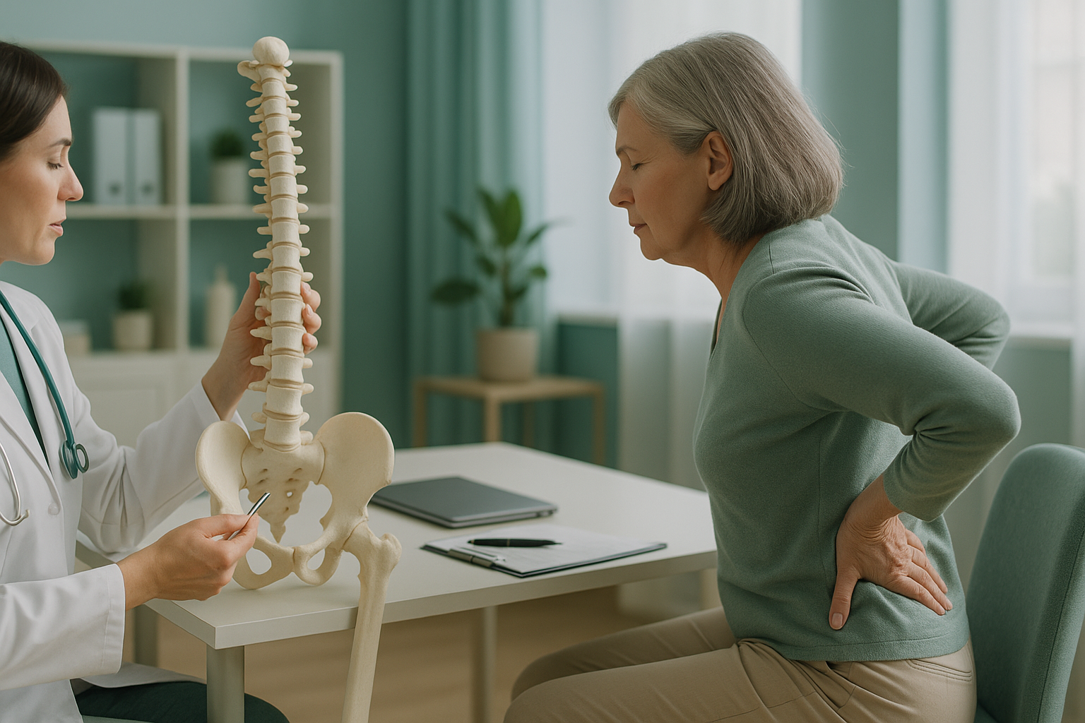
After menopause, oestrogen drops sharply. This speeds up bone loss across the whole skeleton. Bones become thinner, fractures happen more easily, and the spine can slowly change shape. These posture changes often develop slowly. They are easy to miss until pain, height loss or a visible stoop appears.
Key points
- Oestrogen protects bone density. When levels fall at menopause, bone breaks down faster than it rebuilds.
- Weakened vertebrae can slowly compress. This leads to height loss and a greater curve in the spine.
- A DEXA scan measures bone density. A standing EOS scan shows how those changes affect your posture and alignment under load.
- Weight-bearing exercise, enough calcium and vitamin D, and medical advice on HRT all support bone health.
- Knowing early means acting early. Most posture changes are manageable when caught in time.
Why does menopause speed up bone loss?
Oestrogen acts as a brake on the cells that break down old bone. When oestrogen falls during and after menopause, those cells become more active. Bone is removed faster than new bone can be laid down. The result is an overall loss of bone mineral density. This happens most in the first five to ten years after the last period.
This is why the risk of osteoporosis rises sharply in women after menopause. Osteoporosis literally means "porous bones." The outer shell of each bone becomes thinner. The spongy inside becomes more open, like a honeycomb losing its walls.
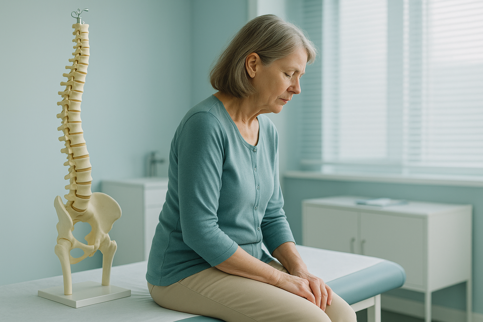
Not every woman gets osteoporosis. But all women lose some bone density after menopause. How fast depends on genetics, lifestyle, body weight, and whether they use hormone replacement therapy (HRT). The key point: even moderate bone loss can change the way the spine holds itself upright.
What are the signs of bone and posture problems after menopause?
Bone loss itself is silent. You cannot feel it happening. The first clue is often a fracture from a minor bump or fall. Or it may be a slow change in posture that someone else notices before you do.
Losing more than a couple of centimetres over time may be a sign that weakened vertebrae are compressing.
A forward stoop that develops slowly, sometimes called a dowager's hump.
A dull, aching pain in the middle or lower back that gets worse with standing or walking.
Many women notice new aches in the hips, knees and shoulders around menopause. This is partly driven by hormonal changes in cartilage and joint fluid.
A note on sudden or severe symptoms. If you have sudden, intense back pain after a fall or minor knock, numbness or weakness in your legs, or loss of bladder or bowel control, get urgent medical help from your doctor or the nearest emergency department. These could point to a fracture or nerve compression that needs checking straight away.
How does bone loss change your posture over time?
When vertebrae weaken, they can compress under your own body weight. This usually happens at the front of the vertebra, making a slight wedge shape. One wedge is hard to notice. Several, stacked up, push the upper spine into a stronger forward curve (kyphosis). This is the classic rounded-shoulder stoop that often develops in later life.
Your body is a connected chain from your feet to your head. So this forward shift causes the body to compensate elsewhere. Your pelvis may tilt to keep your centre of gravity balanced. Your lower back may flatten or arch too much to make up the difference. Your neck pushes forward so your eyes stay level. Pain often appears in these compensating areas, not in the part of the spine that changed first.
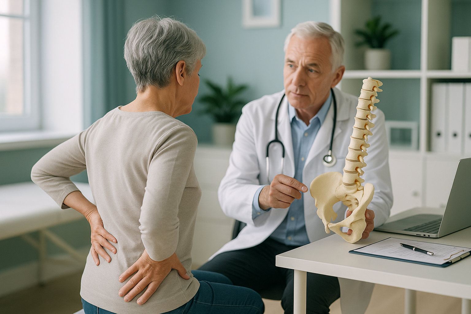
This is why looking at one area on its own can miss the real story. A knee X-ray may look fine. But if your pelvis has tilted to make up for a rounding upper back, your knee carries load unevenly every time you stand or walk.
How are posture changes after menopause assessed?
Two different scans answer two different questions. Both may be useful, depending on your situation.
What it measures: bone mineral density, usually at the hip and lower (lumbar) spine. A DEXA scan tells you how strong your bones are and whether you meet the threshold for osteoporosis or its earlier stage, osteopenia.
When you need it: if your doctor or specialist wants to assess your fracture risk, check how treatment is working, or decide whether to start medicine or HRT. DEXA is the gold standard for measuring bone density.
EOS does not measure bone density and is not a substitute for DEXA.
What it shows: a full-length, weight-bearing image of your skeleton from head to feet, taken while you stand. It shows how your spine curves, whether your pelvis is level, and how your legs and joints are aligned under gravity.
When you need it: if you have back or joint pain, a visible change in posture, height loss, or you want to understand how your body is compensating. The EOS scan uses about 90% less radiation than a standard X-ray, around ten times less.
ScanAlign standing EOS imaging captures your whole skeleton in one scan, standing up, so nothing is hidden by lying down.
The two scans work well together. DEXA tells you about bone quality. A standing EOS scan shows what that bone quality means for the structure of your posture and alignment. If you have already had a DEXA showing low bone density, a standing scan can show whether your vertebrae have started to change shape and how the rest of your body is responding.
When MRI is the better choice. EOS shows bones, joints and alignment. For soft tissue, discs or nerves, MRI is usually the better tool. If your symptoms include nerve pain, tingling or disc-related problems, your specialist may recommend MRI instead, or as well.
It also helps to know that standard lying-down X-rays and MRI scans relax your muscles and take the load off your skeleton. This can hide alignment problems that only show up when you are standing and weight-bearing, which is how you actually spend your day.
What can you do to protect your bones and posture after menopause?
A lot, and the earlier you start, the better. The advice below is general guidance. Your doctor or specialist should shape a plan around your own health history.
Walking, jogging, dancing and resistance training stimulate bone-building cells. Aim for regular activity, ideally on most days.
Both are essential for bone strength. Food, sunlight and supplements (if your doctor advises them) all play a role.
Hormone replacement therapy can slow bone loss and may reduce fracture risk. It is not suitable for everyone, so a one-to-one conversation is important.
A DEXA scan measures bone density. A standing alignment scan shows whether posture changes are already developing. Knowing puts you in control.
If vertebral compression fractures have already happened, a specialist may consider treatments such as kyphoplasty or vertebroplasty. Conditions like osteomalacia (softening of the bones due to vitamin D deficiency) can look like or overlap with osteoporosis. They should be ruled out with blood tests.
Wondering whether your posture has changed? A standing EOS scan at ScanAlign in London can show how your whole skeleton is aligned under your own body weight.
Book a free video consultationIs a standing alignment scan right for you?
A standing EOS scan helps when you want to see how your whole skeleton works together, not just one joint or one area. It is not the right test for every question.
- Have noticed height loss or a developing stoop
- Have back or joint pain that nobody has fully explained
- Want a baseline alignment check after menopause
- Have low bone density on DEXA and want to see the structural picture
- Are thinking about seeing a specialist and want full alignment data
- Need a bone density measurement (DEXA is the right scan for that)
- Have disc, nerve or soft-tissue symptoms (MRI is usually better)
- Have sudden severe pain, weakness, or loss of bladder or bowel control (get urgent medical help)
Before you start a treatment, or change the one you are on, it is worth getting a detailed diagnosis. Knowing whether the cause sits in the pelvis, the legs or the spine changes what will actually help.
Frequently asked questions
No. All women lose some bone density after menopause, but not everyone reaches the threshold for osteoporosis. Genetics, diet, exercise, body weight, and whether you use HRT all affect your own risk.
No. EOS does not measure bone mineral density. A DEXA scan is the correct test for that. EOS shows your skeletal alignment and posture under load. This is a different piece of information that adds to DEXA.
An EOS scan uses about 90% less radiation than a standard X-ray, around ten times less. This makes it suitable for whole-body imaging with very low exposure.
No. ScanAlign is a private, self-pay clinic. You can refer yourself by booking a free video consultation. No referral letter from your doctor is needed. Your report can be shared with your doctor or specialist afterwards.
Research shows that HRT slows bone loss and can reduce the risk of fractures during and after menopause. However, it is not suitable for everyone. The decision should be made with your doctor, based on your full health picture.
Osteoporosis means the bone structure is becoming porous and fragile. Osteomalacia means the bone itself is softening, usually due to severe vitamin D deficiency. They can exist together, and both weaken the skeleton in different ways.
When you lie down, gravity is removed, your muscles relax, and your spine spreads out. Alignment problems that are present when you are upright and weight-bearing may flatten out on a lying-down scan. A standing EOS scan captures your skeleton the way it actually works during your day.
It is a visible rounding at the top of the back, often linked to vertebral compression fractures or increased thoracic kyphosis. It develops slowly and is more common after menopause. You can read more about dowager's hump and how it is assessed.
The price for patients travelling from abroad is confirmed during the free video consultation, which you can use to book a standing posture scan.
Vertebral compression that has already happened cannot usually be fully reversed. But further progression can often be slowed or stopped. Strengthening exercises, treating the underlying bone loss, and addressing alignment can all help manage symptoms and prevent them getting worse.
Understand how your posture has changed
A free video consultation takes about 15 minutes, so you can have it from home before deciding to travel to London. You will speak with a specialist diagnostic radiographer who can explain whether a standing alignment scan is the right next step for you.
Book a free video consultationSources
- Royal Osteoporosis Society. "Osteoporosis: causes and risk factors." theros.org.uk
- NHS. "Menopause: health risks." nhs.uk/conditions/menopause
- Kado DM, Prenovost K, Crandall C. "Narrative review: hyperkyphosis in older persons." Annals of Internal Medicine, 2007;147(5):330-338.
Written by Abbas Dhami (Specialist Diagnostic Radiographer)
This page is general information and not a diagnosis.
ScanAlign is the trading name of CoreMed Solutions Ltd and operates under The Harley Street Hospital's CQC licence.