
How Do Pilates and Movement Instructors Use Alignment Data?
Standing alignment imaging gives Pilates and movement instructors an objective picture of a client's skeletal structure under load. It shows what is structural and what is trainable, helping you programme safely, track real progress, and know exactly when a client needs a medical referral rather than another session.
Key Takeaways
- Alignment data separates structural findings from movement habits you can influence.
- Objective baseline measurements let you demonstrate client progress clearly.
- Knowing when to refer protects your client, your reputation, and your insurance.
- Standing imaging captures the skeleton under gravity, unlike lying-down scans.
- No GP referral is needed for a private standing EOS scan.
Why do Pilates instructors need objective alignment data?
Because visual and palpation-based postural assessment has limits. You can see that a client's shoulder sits higher on one side or that their pelvis appears tilted. But you cannot tell from observation alone whether that asymmetry comes from a bony leg length difference, a spinal curve, a hip joint variation, or simply a muscle imbalance. Those causes require very different programming approaches.
Alignment imaging gives you a baseline measurement of the whole skeleton, taken while your client is standing and weight-bearing. That matters because lying-down imaging, such as a standard X-ray or MRI, relaxes the muscles and can hide asymmetries that only appear under gravity. A standing image shows what their body actually does when it is upright.
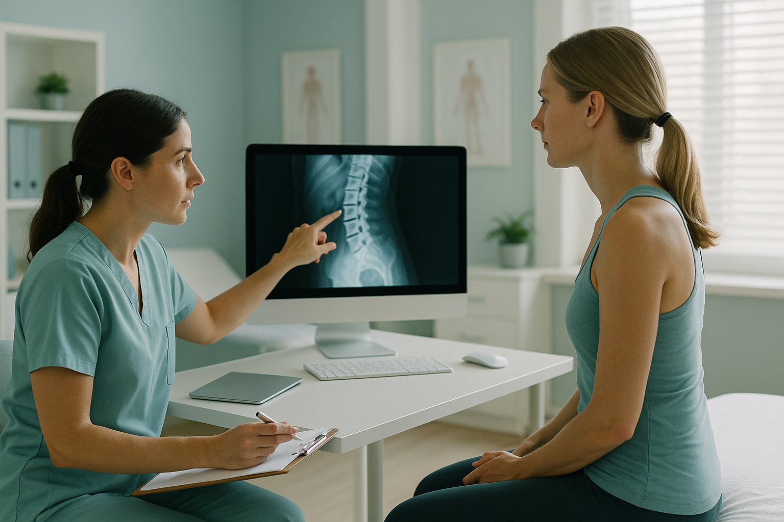
With this data, you can confidently decide which asymmetries will respond to your programming and which ones are structural. That distinction changes everything, from exercise selection to the expectations you set with your client.
How does alignment imaging help you stay within scope of practice?
It draws a clear, documented line between what you can work on and what falls outside your remit. As a movement instructor, your scope of practice covers exercise programming, movement re-education, and motor control. It does not extend to diagnosing medical conditions or treating pathology.
- Programming around a confirmed structural finding
- Tracking changes in movement quality over time
- Sharing imaging reports with a client's physiotherapist or consultant
- Adapting reformer or mat work to suit a known scoliosis
- Interpreting imaging yourself to diagnose a condition
- Telling a client they have a disc problem or joint disease
- Prescribing exercises as a treatment for a medical condition
- Ignoring red flags and continuing to train through pain
When you have a radiologist-reported alignment scan in the client's file, you are not guessing. You are working from documented findings, which is better for the client and better for your professional standing. It also strengthens your insurance position, because you can demonstrate that your programming decisions were informed by clinical data.
When should you refer a Pilates client instead of continuing to train them?
Any time you suspect that what you are seeing goes beyond a movement or muscle issue. Some situations call for imaging or medical assessment before further exercise.
If a client's symptoms consistently increase during or after sessions, something may need investigating before you continue.
If weeks of targeted work produce no change in a tilt or shift, it may be structural rather than muscular.
Working with scoliosis clients is valuable, but you need to know the curve's characteristics before you programme around them.
If a client is approaching or recovering from joint surgery, an up-to-date alignment picture helps you and their surgeon stay on the same page.
A note on soft-tissue concerns. EOS imaging shows bones, joints, and alignment beautifully. For soft-tissue problems such as disc issues, nerve compression, or ligament tears, MRI is usually the better tool. If your client's symptoms suggest a soft-tissue cause, guide them towards MRI rather than an alignment scan.
Important: if a client reports numbness, weakness, loss of bladder or bowel control, or sudden severe symptoms, advise them to seek urgent medical attention through their GP, NHS 111, or A&E. Do not attempt to manage these situations in the studio.
How does standing EOS imaging work for your clients?
ScanAlign standing EOS imaging captures a full-length picture of the skeleton from head to feet in a single standing exposure. Because the client is upright and weight-bearing, the image reveals how their bones, joints, and curves actually behave under gravity, not how they look when relaxed on a table.
The scan shows spinal curves, pelvic position, hip angles, knee alignment, and leg length from a single image. A consultant radiologist reports the findings, so you receive a clinical document rather than a raw image. That report tells you what is structural, giving you the information you need to programme with confidence.
EOS uses about 90% less radiation than a standard X-ray, around ten times less. The scan takes only seconds and requires no preparation. Your client stands still, fully clothed (metal-free clothing), and walks out immediately.
The body is a connected chain. A structural issue at the hip can cause the spine to compensate, and that compensation might show up as shoulder tension or lower-back stiffness. Whole-body standing imaging captures that entire chain in one go, which is exactly why it is so useful for instructors who think in terms of whole-body movement patterns.
How do you refer a client for a standing alignment scan?
The process is simple. No GP referral is needed. You can either suggest that your client books directly, or you can register to refer clients through a simple professional referral pathway.
You notice an asymmetry, a plateau, or a concern that falls outside your scope. You and your client agree that objective data would help.
Either refer a client through the professional pathway, or ask them to book a free video consultation themselves. No paperwork, no GP letter.
Your client attends 19 Harley Street for their standing EOS scan. A consultant radiologist reviews the images and produces a detailed report.
With the report in hand, you know what is structural and what you can influence. You can adapt your programming, set realistic goals, and track objective progress over time.
Want to build alignment imaging into your client assessment process? Register as a referring professional and receive reports directly.
Register to refer clientsFrequently asked questions
No. Pilates instructors, movement coaches, personal trainers, and other movement professionals can all refer clients for a private standing EOS scan. No GP letter is required.
With the client's consent, the report can be shared with you as the referring professional. This allows you to integrate the findings into your programming.
Yes. EOS uses about 90% less radiation than a standard X-ray, around ten times less. It is one of the lowest-dose skeletal imaging methods available.
Pricing details are available on the ScanAlign FAQs page. A free video consultation is the first step, during which the team will discuss costs and what the scan involves.
A standing alignment scan shows the full picture of a scoliosis under gravity. Knowing the curve's characteristics helps you programme reformer or mat work that respects the client's structure rather than fighting it.
They serve different purposes. EOS shows bones, joints, and alignment. MRI is the better tool for soft-tissue problems such as discs, nerves, or ligaments. Some clients may benefit from both.
The scan itself takes only seconds. Your client should allow a short time for check-in and positioning. There is no special preparation needed.
Yes. A follow-up scan after a period of training gives you an objective comparison, showing whether postural changes are occurring at a skeletal level. This is far more compelling than visual observation alone.
Having clinical imaging to support your programming decisions strengthens your position. It shows you acted on documented evidence rather than assumption, which is good practice from both a clinical and insurance perspective.
ScanAlign is based at 19 Harley Street, London, operating under The Harley Street Hospital's CQC licence.
Ready to add alignment data to your client assessments?
A free video consultation is the first step. We will discuss how standing imaging fits into your practice and answer any questions.
Book a free video consultationSources
- Deschênes S, Charron G, Beaudoin G, et al. "Diagnostic imaging of spinal deformities: reducing patients radiation dose with a new slot-scanning X-ray imager." Spine, 2010; 35(9): 989-994.
- The Chartered Society of Physiotherapy. "Scope of practice guidance for physiotherapists and support workers." CSP, 2024.
- Pilates Method Alliance. "Scope of practice for Pilates professionals." PMA position paper, 2015.
Written by Abbas Dhami (Specialist Diagnostic Radiographer)
This page is general information and not a diagnosis.
ScanAlign is the trading name of CoreMed Solutions Ltd and operates under The Harley Street Hospital's CQC licence.