
Are Bow Legs Normal in Toddlers, and When Do They Straighten?
Yes, bow legs in toddlers are almost always normal. Most babies are born with naturally curved legs from being tucked up in the womb. The bowing usually corrects on its own by around age three to four. If the curve is severe, getting worse, or only on one side, a specialist assessment can help rule out rarer conditions.
Key takeaways
- Bow legs (genu varum) are a normal part of development in babies and toddlers.
- Most children's legs straighten naturally between the ages of two and four.
- Warning signs include bowing that worsens after age two, affects only one leg, or causes pain or limping.
- A standing, weight-bearing scan shows alignment under real-world conditions, which lying-down imaging cannot replicate.
- ScanAlign offers paediatric imaging at ScanAlign with about 90% less radiation than a standard X-ray.
What are bow legs in a toddler?
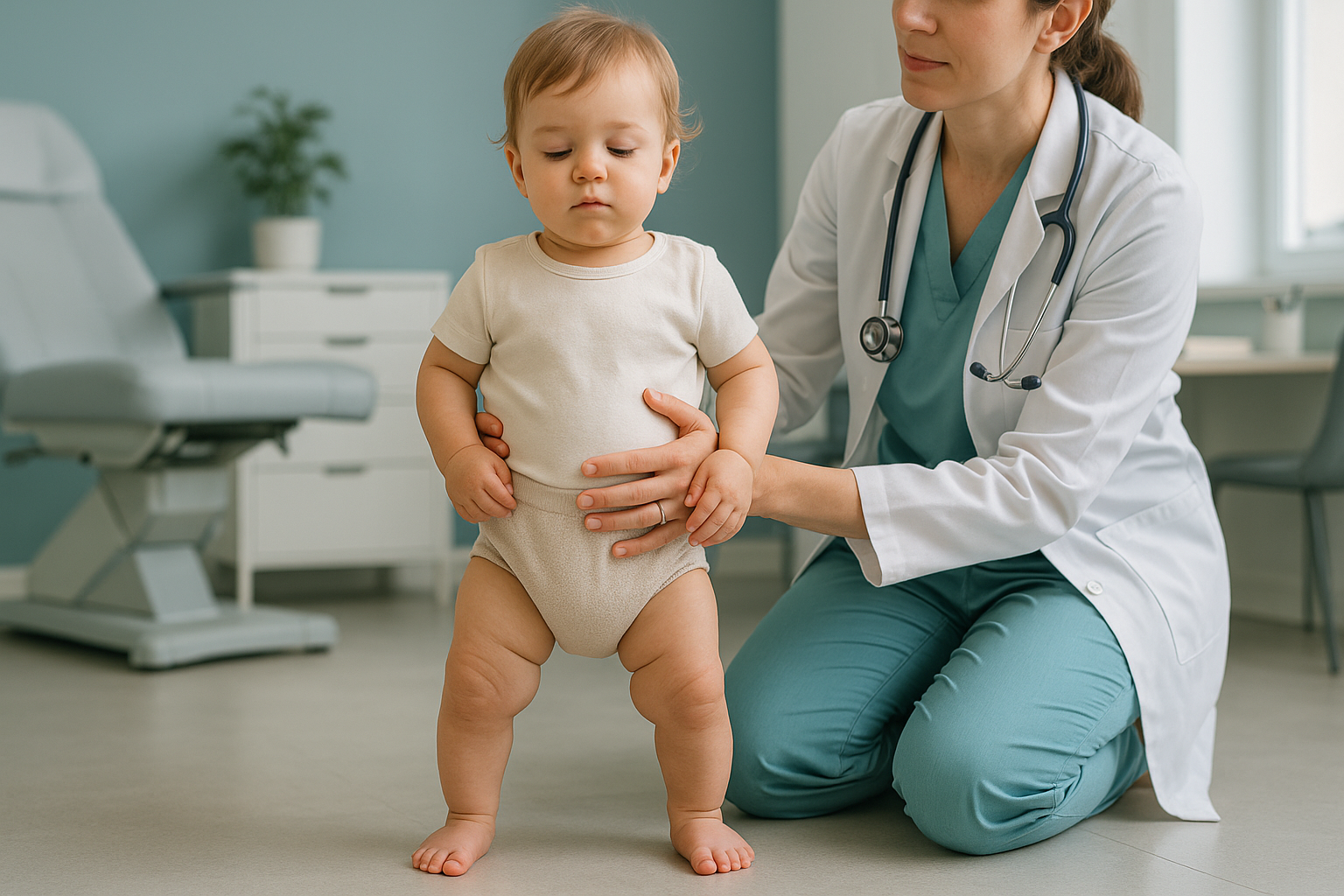
Bow legs, known medically as genu varum, describes a visible outward curve at the knees when a child stands with their ankles together. The knees stay apart, creating a gap. In medical terms, the distance between the inner edges of the knees is called the intercondylar distance.
Nearly all newborns have some degree of leg bowing. This is called physiological bowing, and it happens because of the baby's folded position inside the womb during the last weeks of pregnancy. It is not a sign of a problem. It is simply how the bones develop before birth.
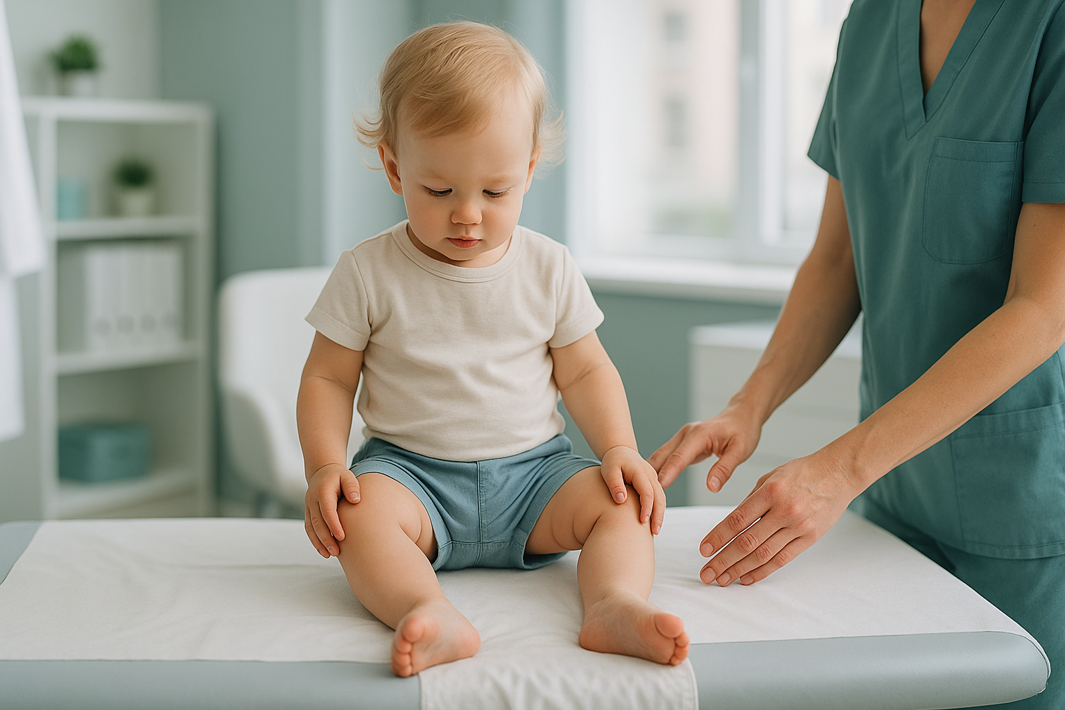
As your child starts to walk and bear weight through their legs, the bones gradually remodel. This is a normal, predictable process. Many children then pass through a brief phase of knock knees before the legs settle into their adult alignment.
When do bow legs straighten in children?
Most bow legs straighten on their own between the ages of two and four. By the time a child is around 18 to 24 months old, the curve typically starts to reduce. By age three to four, the legs are usually straight or even slightly knock-kneed, which is also normal at that stage.
The process is gradual. You might not notice changes week to week, but over several months the gap at the knees closes. No treatment is usually needed. Your GP or health visitor may suggest watchful waiting, which simply means keeping an eye on the shape over time.
If the bowing has not started to improve by age two, or if it is clearly getting worse rather than better, that is a good reason to seek a specialist opinion.
What signs suggest bow legs might not be normal?
In most toddlers, bow legs need no intervention at all. However, certain features can suggest that the bowing is not purely physiological and may benefit from further assessment.
What causes bow legs in babies and toddlers?
The most common cause is simply normal development. In a small number of children, the bowing has a different origin that may need monitoring or treatment.
Other rare causes include skeletal dysplasia and previous fractures near the knee. Your GP can usually tell the difference between normal bowing and something that needs investigation based on your child's age, the severity of the curve, and whether it is changing over time.
How does a standing scan help assess bow legs?
A standing, weight-bearing scan captures your child's leg alignment exactly as it works in real life, while gravity is pulling on the joints and bones. This matters because lying-down imaging relaxes the muscles and can make the legs look straighter than they really are, potentially hiding the true extent of the bowing.
EOS imaging, available through ScanAlign, low dose standing imaging, takes a full-length image of both legs in a single standing exposure. This gives the clinician a clear picture of the overall alignment from hips to ankles, not just a small section of one bone. Read more about bow legs and knock knees on a standing scan.
Importantly, an EOS scan uses about 90% less radiation than a standard X-ray, around ten times less. For children, reducing radiation exposure is a real priority, and low-dose imaging makes parents and clinicians more comfortable with the assessment.
If your child's GP or specialist has recommended imaging, or if you would like a clearer picture of their leg alignment, you can book a paediatric assessment at our Harley Street clinic.
Book a free video consultationWhen is MRI better? EOS shows bones, joints and alignment clearly. If your child's specialist suspects a soft-tissue problem, a cartilage issue, or needs to look at the growth plates in fine detail, MRI is usually the better tool. Your clinician will advise which scan is right.
Frequently asked questions about bow legs in toddlers
If your child's legs are still bowing at age two or if the curve is getting worse rather than better, it is worth speaking to your GP. Most bowing resolves by age three to four without any intervention.
Not usually. Bow legs in babies are almost always physiological. Rickets is uncommon in the UK but can occur with low vitamin D levels. Your GP can check with a simple blood test if there is concern.
Yes. Physiological bowing, which accounts for the vast majority of cases, corrects on its own as the child grows. Watchful waiting is the standard approach.
Normal physiological bowing does not cause pain. If your child is experiencing pain, limping, or seems reluctant to walk, speak to your GP as this could point to a different cause.
Blount's disease is a growth plate disorder affecting the inner part of the upper shin bone (tibia). It causes progressive bowing that does not improve with age and typically needs specialist management.
No. You can self-refer. We offer a free video consultation first so our specialist radiographer can check that a scan is appropriate for your child's situation.
Yes. EOS uses about 90% less radiation than a standard X-ray, around ten times less. This makes it a very low-dose option, which is especially important for growing children.
The scan itself is very quick. Your child will need to stand still for a short time. Our team are experienced with young children and will help make the process as smooth as possible. We discuss suitability in the free video consultation beforehand.
We do not list prices on individual pages. You can find current pricing and what is included on our paediatric imaging at ScanAlign page, or discuss costs during your free video consultation.
You receive a detailed report from a specialist radiologist. This can be shared with your child's GP, paediatrician, or orthopaedic surgeon to guide any next steps. ScanAlign is a diagnostic imaging clinic and does not provide treatment.
Concerned about your child's leg alignment?
A free video consultation with our specialist radiographer lets you discuss your child's situation, ask questions, and find out if a standing scan is the right next step.
Book a free video consultationSources
- NHS. "Bow legs." nhs.uk. Accessed 2025.
- American Academy of Orthopaedic Surgeons. "Bow Legs (Genu Varum)." OrthoInfo. Accessed 2025.
- Sabharwal S. "Blount disease." Journal of Bone and Joint Surgery, 91(7), 2009.
Written by Abbas Dhami (Specialist Diagnostic Radiographer)
This page is general information and not a diagnosis.
ScanAlign is the trading name of CoreMed Solutions Ltd and operates under The Harley Street Hospital's CQC licence.