
Knock Knees: What Causes Them in Adults and How Are They Assessed?
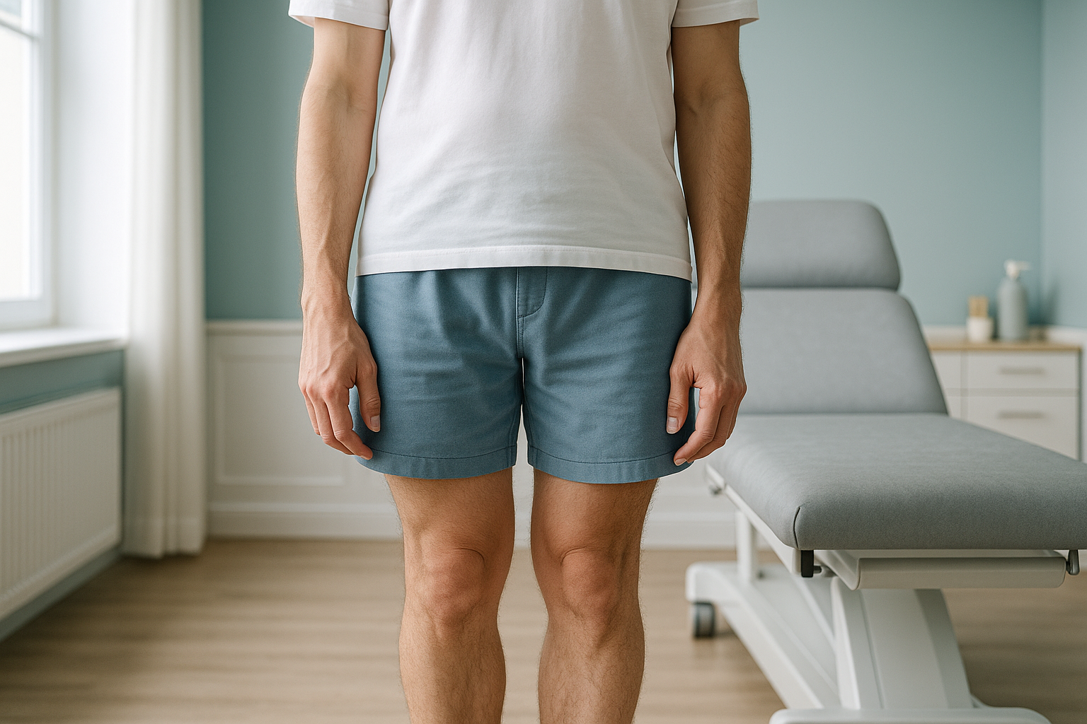
Knock knees (genu valgum) is a leg alignment where the knees angle inward while the ankles stay apart. In adults it can strain the knees, hips and lower back. A standing, weight-bearing scan shows the true alignment under load, something a lying-down scan can miss, so the right treatment plan can begin.
Key takeaways
- Knock knees in adults are not just cosmetic. They change how force travels through the whole body.
- Common causes include previous injury, arthritis, obesity and muscle imbalance.
- Pain often appears in the knee, but the root cause may sit in the hip, ankle or pelvis.
- A standing whole-body scan captures alignment under real weight-bearing conditions.
- ScanAlign provides diagnostic imaging only. It does not treat or correct knock knees.
Most people have heard of knock knees in young children, where it is a normal stage of growth. But when the same alignment persists or develops in adults, it can cause real problems. Your knees sit at the centre of a chain that runs from your feet to your spine. When they angle inward, that chain is disrupted, and pain can show up in places you would not expect.
What does knock knee alignment look and feel like?
Knock knees are visible when your knees touch or overlap but your ankles stay apart when you stand with your legs straight. Beyond the visible angle, you may notice several day-to-day effects.
The inward angle overloads the outer compartment and stretches the inner ligaments, causing aching around the knee.
The pelvis tilts to compensate for the knee angle, which can create stiffness or pain higher up in the chain.
The soles of your shoes may wear down more on the inner edge, reflecting how your weight shifts inward as you walk.
Running, squatting or climbing stairs may feel unstable or painful because the joint is loaded unevenly.
What causes knock knees in adults?
In adults, knock knees usually develop because of a structural or biomechanical change rather than normal growth. The most common causes include the following.
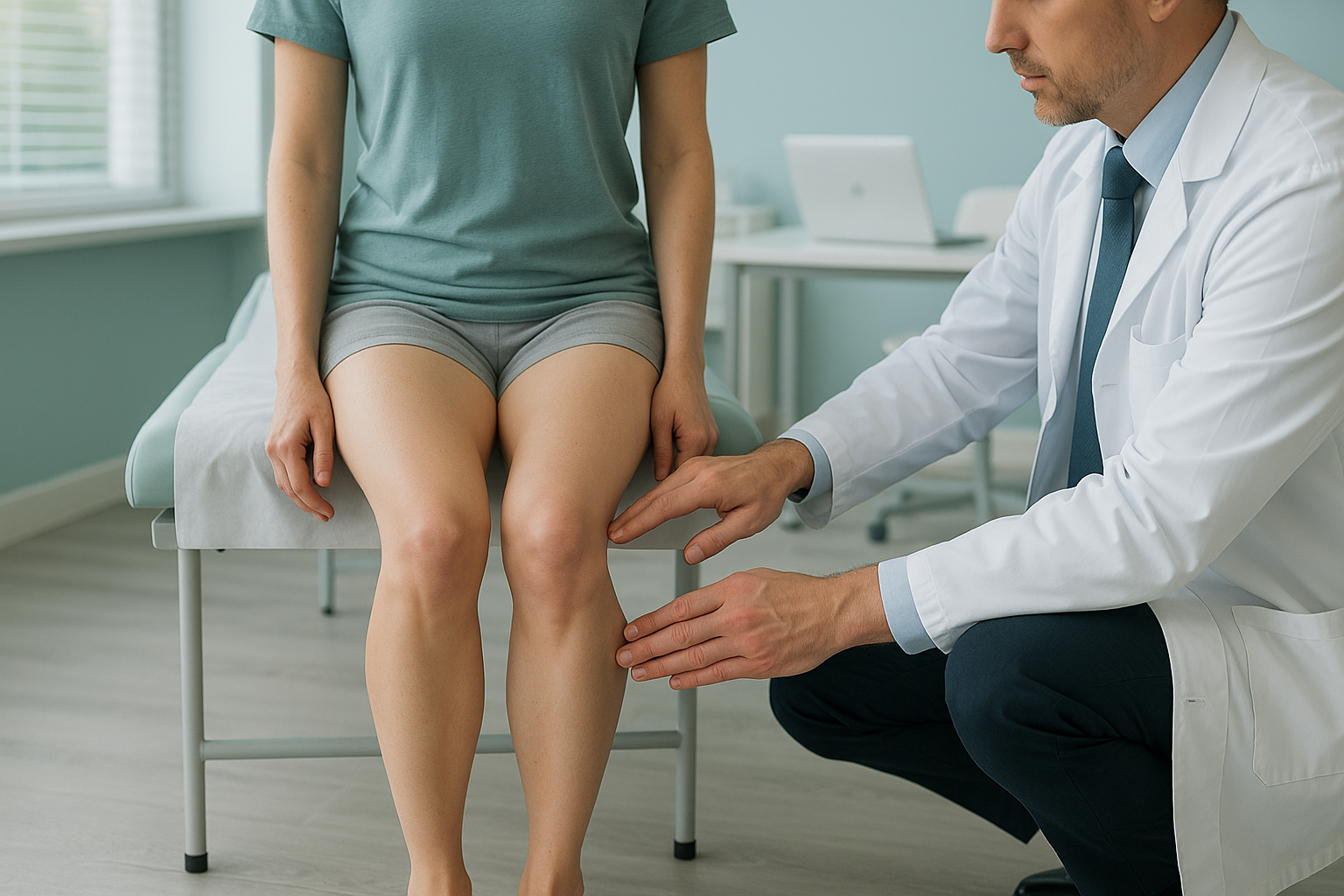
Cartilage loss on one side of the knee changes the joint angle over time.
A break near the knee that heals at a slight angle can gradually shift alignment.
Extra body weight increases inward force at the knee, worsening the valgus angle.
Weak outer hip muscles let the thigh rotate inward, pulling the knee with it.
Other possible causes include rickets (vitamin D deficiency affecting bone), rheumatoid arthritis, and ligament damage. Sometimes more than one factor combines.
How do knock knees affect the rest of your body?
Your body works as a connected chain from your feet to your head. When the knees angle inward, every joint above and below has to adjust. This is called compensation, and it is why you might feel pain far from your knees.
The ankles may roll inward (over-pronation) to stay flat on the ground. The hips may rotate to keep you balanced. The pelvis can tilt, and the lower back curves differently to keep your head level. A single-area scan of the knee might show no obvious damage, yet the real issue is the way every joint interacts under your full body weight.
Want to understand how your whole alignment connects? Explore how standing imaging assesses the knee and leg.
How is knock knee alignment assessed when standing up?
True leg alignment can only be seen when you are upright and bearing weight. Lying-down imaging relaxes your muscles and can flatten out the angles that cause your symptoms. A standing, whole-body EOS scan captures your skeleton from head to toe in a single upright image, showing the exact relationship between your feet, knees, hips, pelvis and spine under real-world load.
EOS uses about 90% less radiation than a standard X-ray, around ten times less. You simply stand still for about 20 seconds while the system produces a full-length image. Your clinician then receives a detailed picture of your alignment, not just one joint in isolation.
When MRI is the better choice: EOS shows bones, joints and alignment clearly. For soft-tissue problems like cartilage tears, ligament damage or nerve issues, MRI is usually the better tool. Your clinician can advise which scan you need.
What are the general approaches to managing knock knees?
Management depends entirely on the cause and severity, which is why accurate imaging matters first. Generally, clinicians may consider the following options.
- Physiotherapy and targeted exercises. Strengthening the muscles around the hip and knee can improve how the joint is held, especially when muscle imbalance is involved.
- Orthotic insoles. Custom insoles may help correct foot position, which in turn can reduce inward knee force.
- Weight management. Reducing excess load on the knees can slow the worsening of alignment.
- Surgery. In more severe or progressive cases, procedures such as osteotomy (realigning the bone) or knee replacement may be considered by a specialist.
ScanAlign does not provide treatment or therapy. It provides the standing diagnostic imaging that helps your clinician or surgeon plan the right next step. For information on what to expect and costs, visit the FAQs page.
Knock knees may also affect hip alignment. Read more about how standing imaging assesses hip alignment.
When to seek urgent help
If you experience sudden knee swelling, inability to bear weight, numbness, weakness in the leg, or loss of bladder or bowel control, seek urgent medical attention. Contact your GP, call 111, or go to A&E.
Frequently asked questions about knock knees
In adults, the most common causes are osteoarthritis, previous fracture or injury near the knee, obesity, muscle imbalance, and occasionally conditions like rheumatoid arthritis or vitamin D deficiency. Often several factors combine over time.
A slight inward angle is within the normal range for many adults. It becomes a concern when it causes pain, limits activity, or shows signs of worsening. A standing alignment assessment helps clarify whether the angle is within normal limits or needs attention.
Targeted strengthening of the hip and thigh muscles can help improve how the knee is held, particularly when muscle weakness is a contributing factor. However, if the bone itself has changed shape, exercises alone may not fully correct the angle. A physiotherapist can guide an appropriate programme after imaging has identified the cause.
When you lie down, gravity is no longer compressing your joints and your muscles relax. This can flatten out the inward angle and hide the true extent of the misalignment. A standing, weight-bearing scan captures alignment as it actually is when you are upright and loading your joints.
An EOS scan uses about 90% less radiation than a standard X-ray, around ten times less. This makes it a very low-dose option for full-body standing imaging.
No. ScanAlign is a private, self-pay clinic and you can self-refer. You start with a free video consultation to check the scan is appropriate for you.
For the latest pricing and what is included, please visit the FAQs and cost page.
Yes. The inward knee angle changes how force travels up through the hips and pelvis. The lower back often adjusts its curve to keep you balanced. This compensation can create pain in areas that seem unrelated to the knees.
Knock knees (genu valgum) means the knees angle inward and the ankles sit apart. Bow legs (genu varum) is the opposite, where the knees curve outward and the ankles touch. Both affect alignment and loading of the joints differently.
You stand in the EOS scanner for about 20 seconds while it captures a full-length image of your skeleton from head to toe. There is no enclosed tunnel and no need to change into a gown in most cases. Your results are then reviewed and a report is provided for you or your clinician.
Understand your alignment, not just your symptoms
A free video consultation with our team will help you decide whether a standing whole-body scan is the right next step.
Book a free video consultationLearn more about how EOS whole-body standing imaging works.
Sources
- NHS. "Knock knees." nhs.uk. Accessed 2024.
- Sharma L, Song J, Felson DT, et al. "The role of knee alignment in disease progression and functional decline in knee osteoarthritis." JAMA. 2001;286(2):188-195.
- Illés T, Somoskeöy S. "The EOS imaging system and its uses in daily orthopaedic practice." International Orthopaedics. 2012;36(7):1395-1402.
Written by Abbas Dhami (Specialist Diagnostic Radiographer)
This page is general information and not a diagnosis.
ScanAlign is the trading name of CoreMed Solutions Ltd and operates under The Harley Street Hospital's CQC licence.