

What Is Congenital Scoliosis, and How Is It Monitored?
Congenital scoliosis is a sideways curve of the spine caused by vertebral anomalies that form before birth. Unlike other types of scoliosis, the curve is present from day one. Monitoring is essential because growth can make the curve worse. Low-dose standing imaging helps specialist teams track changes over time with less radiation exposure.
Key takeaways
- Congenital scoliosis is caused by vertebrae that did not form or separate properly during early pregnancy.
- It is not caused by posture, carrying bags, or anything the parent did.
- Monitoring is usually led by a specialist centre and may continue through adulthood.
- Repeated imaging means cumulative radiation matters. EOS technology uses about 90% less radiation than a standard X-ray, around ten times less.
- ScanAlign works alongside your specialist team, never as a replacement.
What causes congenital scoliosis?
Congenital scoliosis happens when one or more vertebrae do not develop normally in the first six weeks of pregnancy. The result is a spine that curves to one side because its building blocks are uneven. This is different from adolescent or degenerative scoliosis, which develop later in life for different reasons.
The exact cause is usually unknown. It is not linked to anything the mother ate, did, or was exposed to. In most cases, there is no family history. However, because vertebrae form at the same time as other organs, some people with congenital scoliosis are also screened for heart or kidney differences.
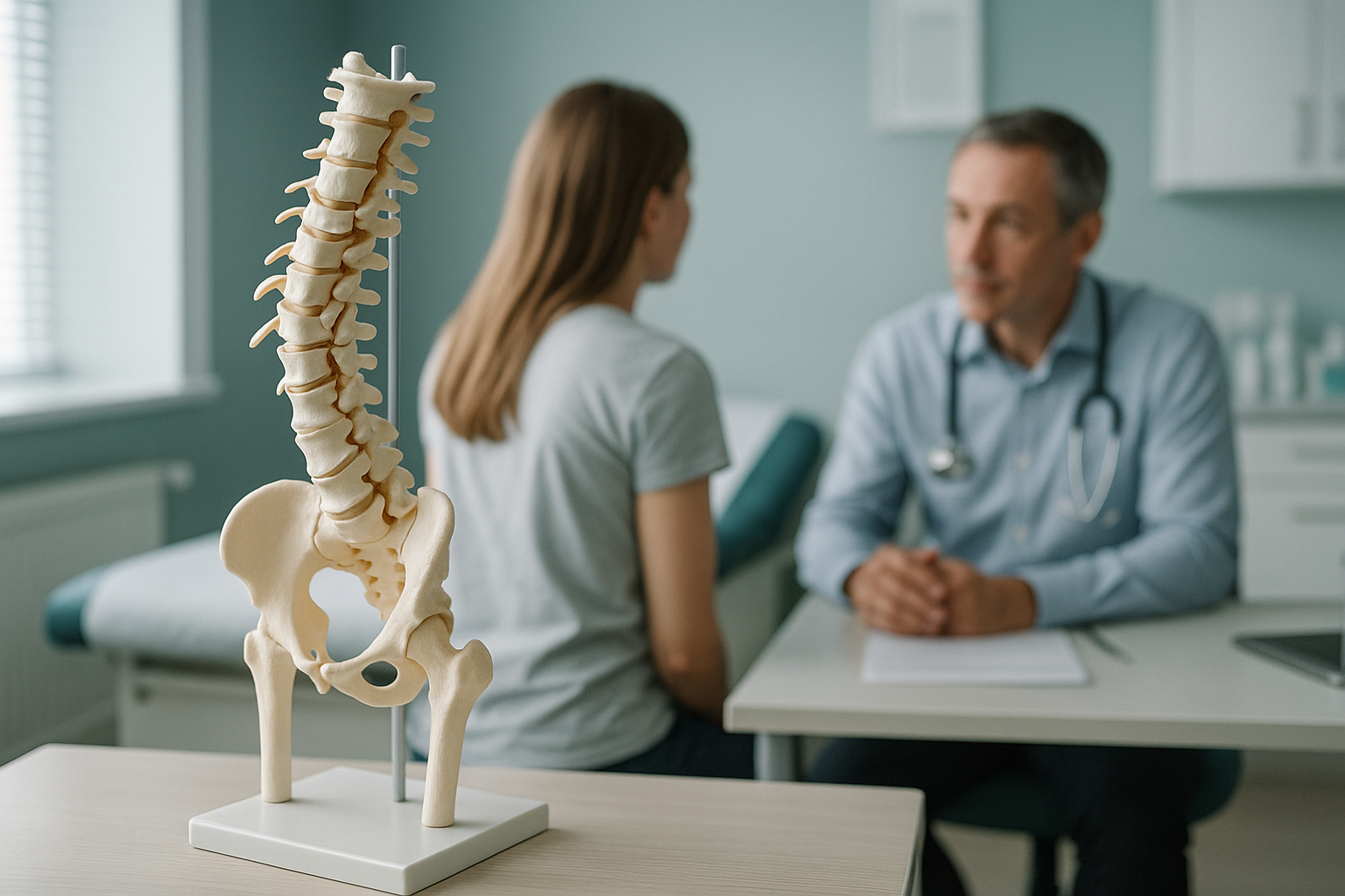
What are the main types of vertebral anomaly?
Vertebral anomalies in congenital scoliosis fall into two broad groups, and some people have a mix of both.
Part of a vertebra does not form at all. The most common example is a hemivertebra, where only one side of the bone develops, creating a wedge shape that pushes the spine sideways.
Two or more vertebrae do not separate properly and are fused together on one side. This tethers growth on that side, so the curve worsens as the child grows.
Some spines have a combination of malformed and fused vertebrae. Mixed anomalies can be harder to predict and often need more frequent imaging.
The type, location, and number of anomalies all influence whether the curve is likely to stay stable or progress during growth. That is why ongoing monitoring matters so much.
What are the signs of congenital scoliosis?
Many congenital curves are noticed in infancy or early childhood, but milder cases can go undetected until a growth spurt makes the curve more visible. Some adults discover the condition only when they are investigated for back pain or stiffness.
One shoulder sitting higher than the other, or the waistline looking asymmetrical.
A noticeable curve when bending forward, or one side of the ribcage appearing more prominent.
Fused or malformed vertebrae can limit flexibility, sometimes causing stiffness or aching that builds gradually over years.
If you notice sudden weakness, numbness, or loss of bladder or bowel control, these are red-flag symptoms. Please seek urgent medical attention from your GP, call 111, or go to A&E straight away.
How is congenital scoliosis monitored over time?
Monitoring is usually led by a specialist spinal centre. The goal is to track whether the curve stays stable or gets worse, especially during periods of rapid growth. Most monitoring plans include regular clinical examinations and imaging at intervals set by the specialist team.
Depending on the type and severity of the curve, the specialist may recommend observation only, bracing in certain situations, or surgery such as growing rods or spinal fusion. ScanAlign does not provide treatment. We offer the imaging that supports your specialist team's decisions.
Why cumulative radiation dose matters
Because congenital scoliosis often requires imaging over many years, the total radiation exposure adds up. Every scan is a small dose, but over dozens of visits those doses accumulate. This is where low-dose imaging technology becomes especially valuable. Using about 90% less radiation than a standard X-ray, around ten times less, helps keep the cumulative dose as low as reasonably possible.
Concerned about cumulative dose from years of monitoring? Ask your specialist whether a low-dose standing scan could be used for your next check. You can also book a monitoring scan and share the results directly with your clinical team.
How does standing imaging help with congenital scoliosis?
Standing imaging captures the spine under real-world conditions, with gravity and your body weight acting on the curve. When you lie down for a conventional X-ray or MRI, the muscles relax and the curve can look less severe than it actually is. A standing, weight-bearing scan shows the curve as your body truly experiences it.
What it shows: The full spine and pelvis in a single upright image. Weight-bearing alignment, compensatory curves, and how the pelvis and hips respond to the spinal anomaly.
Why it matters for congenital scoliosis: Your body compensates for a structural curve by adjusting posture elsewhere. Standing imaging reveals those compensations clearly, giving your specialist the complete picture.
What it shows: Bone and soft-tissue detail, but with the spine unloaded. Muscles relax, and gravity is removed from the equation.
The limitation: The curve may appear smaller than it is in daily life, which can understate the true alignment picture.
ScanAlign, low dose standing imaging uses EOS technology to capture a full-length image of your skeleton while you stand naturally. The scan takes only a few seconds and uses about 90% less radiation than a standard X-ray, around ten times less. For paediatric imaging at ScanAlign, this low dose is especially important given the number of scans a young person with congenital scoliosis may need over the years.
When is MRI the better choice? EOS shows bones, joints, and alignment beautifully. For soft tissue, discs, the spinal cord, or nerves, MRI is usually the better tool. Many people with congenital scoliosis need both types of imaging at different stages. Your specialist team will advise which is right for each check.
Is a standing alignment scan right for you?
A low-dose standing scan can be a useful addition to your monitoring plan, but it is not the right choice for everyone.
- Have an established congenital scoliosis diagnosis and want to reduce cumulative radiation from monitoring
- Are an adult whose curve has not been reassessed in years
- Need a full-length standing image to share with a surgeon or physiotherapist
- Want to see how your whole skeleton compensates for the curve
- Your specialist centre's monitoring programme
- MRI when the spinal cord or nerves need to be assessed
- Cardiac or renal screening for associated anomalies
- Emergency assessment if you have sudden neurological symptoms
Frequently asked questions about congenital scoliosis
Congenital scoliosis is caused by vertebrae that formed abnormally before birth. Idiopathic scoliosis develops later, usually during adolescence, and its cause is unknown. The two conditions behave differently and are managed differently.
It can. Most progression happens during childhood growth spurts, but some curves continue to change slowly in adulthood, particularly if the anomaly creates ongoing imbalance. Periodic imaging helps your specialist spot any gradual change.
In most cases, no. Congenital scoliosis usually occurs without a family history. Researchers have identified some genetic links, but the condition is generally considered sporadic rather than inherited.
A hemivertebra is a vertebra where only one side developed fully, giving it a wedge or triangular shape. It is the most common type of failure of formation and typically causes the spine to tilt to one side.
The vertebrae, kidneys, and heart all begin to form at a similar stage of early pregnancy. A vertebral anomaly can sometimes occur alongside differences in these organs. Screening for associated anomalies is a standard part of the initial workup.
An EOS scan uses about 90% less radiation than a standard X-ray, around ten times less. This is especially important for anyone who needs repeated imaging over many years, where cumulative dose is a concern.
No. You can self-refer. We recommend starting with a free video consultation so we can understand your situation, confirm the scan is appropriate, and make sure the results will be useful to your specialist team.
No. ScanAlign is a diagnostic imaging clinic. We provide the images, but your specialist centre manages your ongoing care, including clinical assessments, treatment decisions, and multidisciplinary support. We work alongside your team, not instead of them.
We do not list prices on individual pages. You can find current pricing and what is included on our frequently asked questions page.
Bring any previous imaging or reports if you have them, along with details of your specialist team so we can share results with them directly if you wish. Wear comfortable clothing without metal fastenings if possible.
Want to discuss whether a standing scan fits your monitoring plan?
A short video call with our specialist radiographer costs nothing and carries no obligation. We can review your history and advise whether EOS imaging would add value for your team.
Book a free video consultationWritten by Abbas Dhami (Specialist Diagnostic Radiographer)
Sources
- Hedequist D, Emans J. Congenital scoliosis: a review and update. Journal of Pediatric Orthopaedics. 2007;27(1):106-116.
- Scoliosis Research Society. Congenital scoliosis overview. srs.org (accessed 2024).
- NHS. Scoliosis: overview, diagnosis and treatment. nhs.uk (accessed 2024).
This page is general information and not a diagnosis.
ScanAlign is the trading name of CoreMed Solutions Ltd and operates under The Harley Street Hospital's CQC licence.