
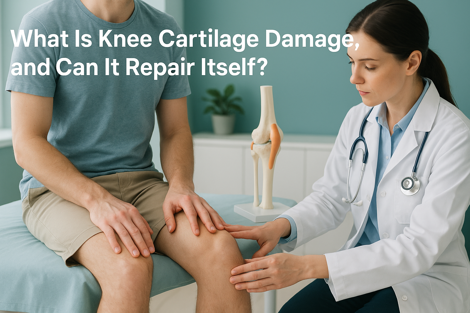
What Is Knee Cartilage Damage, and Can It Repair Itself?
Knee joint cartilage damage is a breakdown of the smooth, protective tissue covering the ends of your thigh bone and shin bone. Because articular cartilage has almost no blood supply, it struggles to heal on its own. Over time, untreated damage can progress to arthritis. Understanding the load your knee carries while standing is key to managing it well.
Key takeaways
- Articular cartilage cushions your knee joint but has very limited ability to repair itself.
- Pain, swelling, catching and stiffness are the most common symptoms of cartilage damage.
- Alignment matters. If your leg's mechanical axis is off, one side of your knee takes more load, accelerating the wear.
- MRI grades the cartilage itself. A standing scan shows how your alignment distributes weight through the damaged area.
- Early assessment gives you more options, from targeted strengthening to surgical repair, before the damage progresses.
What is articular cartilage and why does it matter?
Articular cartilage is a thin layer of firm, slippery tissue that covers the ends of the bones inside your knee. It lets your joint glide smoothly and absorbs shock when you walk, run or climb stairs. Think of it like the non-stick coating on a pan. When it is intact, everything moves freely. When it wears through, bone grinds on bone.
Unlike most tissues in your body, articular cartilage has almost no blood supply. Blood carries the repair cells and nutrients that heal an injury. Without that supply, even a small area of damage tends to stay damaged, or slowly get worse.
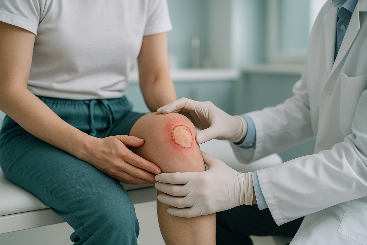
A chondral defect is the clinical name for an area where the cartilage has cracked, thinned or worn away entirely. A related condition, chondromalacia patella, describes softening of the cartilage behind the kneecap specifically.
What does knee cartilage damage feel like?
The symptoms depend on how much cartilage has been lost and where in the joint the damage sits. Some people notice problems after a specific injury. Others find the discomfort creeps in gradually over months or years.
Often felt deep inside the joint, worse during or after activity like stairs or squatting.
The knee may puff up after exercise or at the end of a long day on your feet.
A loose flap of cartilage can catch during movement, making the knee feel like it sticks or gives way.
The knee may feel stiff when you first get up, then loosen off after a few minutes of gentle movement.
A note on sudden symptoms. If you experience sudden, severe knee pain with significant swelling, locking you cannot release, or the knee completely giving way under you, see your GP, call 111, or attend A&E promptly. These can indicate a more serious injury that needs urgent assessment.
What causes knee cartilage damage?
Cartilage damage can start with a single event or build up slowly. Here are the most common causes.
A twist, fall or impact can crack or shear cartilage away from the bone beneath.
Years of repetitive load, especially with poor alignment, slowly thins the cartilage surface.
If the mechanical axis of the leg is tilted, one compartment of the knee takes more than its share of load.
A torn meniscus or ligament reconstruction can change how load is distributed, speeding up cartilage wear elsewhere.
Does cartilage grow back on its own?
In most cases, no. Articular cartilage has very limited ability to repair itself. Small, shallow defects may stabilise and cause few symptoms if the joint is well aligned and the surrounding muscles are strong. But once the damage reaches the subchondral bone beneath, the body's repair attempt produces fibrocartilage, a rougher, weaker tissue that does not perform as well as the original surface.
Left unmanaged, cartilage damage tends to progress. The damaged area spreads, the joint space narrows, and the condition gradually becomes osteoarthritis. That is why early assessment matters. The more cartilage you still have, the more options are available to protect it.
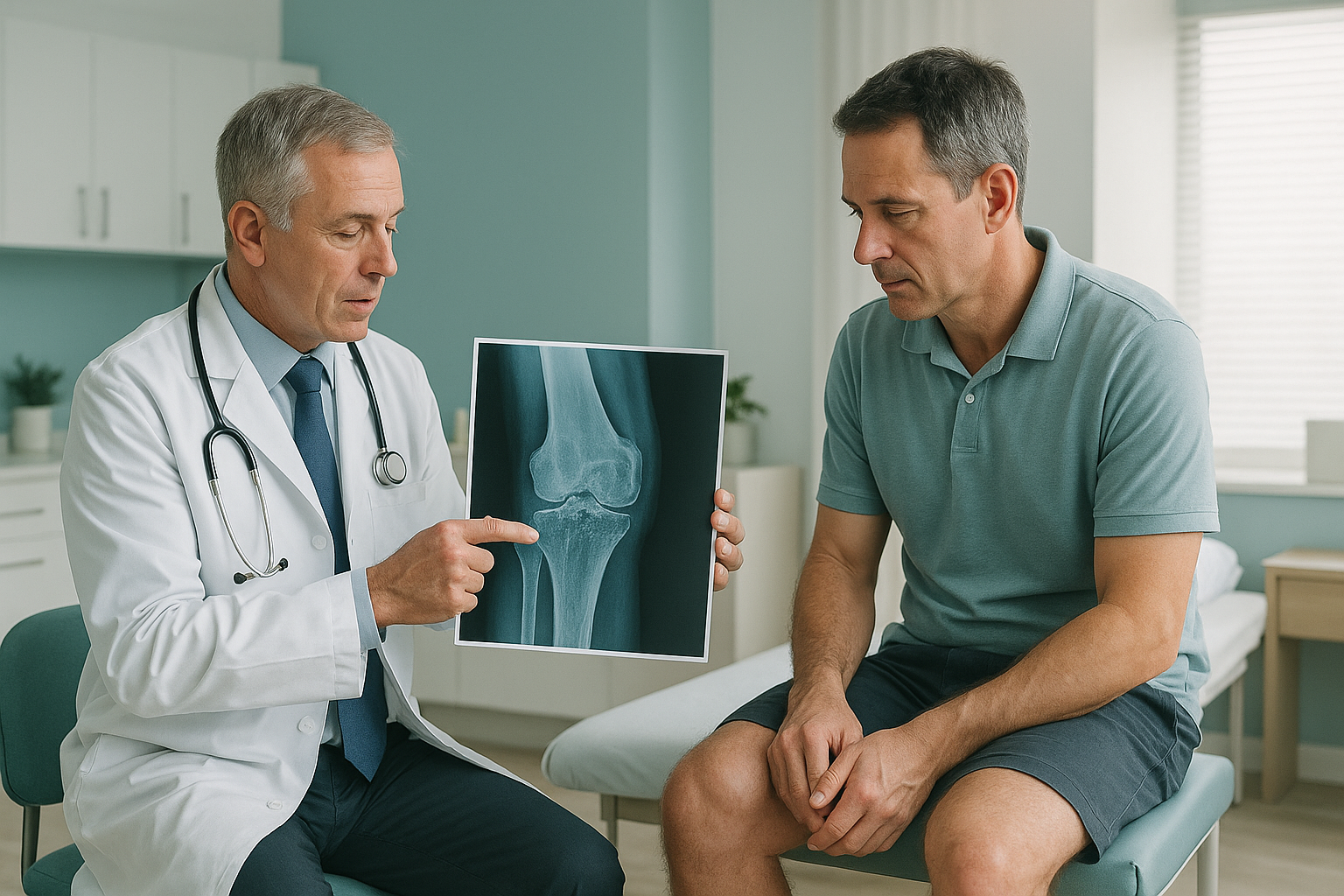
How does a standing scan show what lying-down imaging misses?
A standing, weight-bearing scan captures your knee under real load, the same load it carries every time you take a step. When you lie down for a standard X-ray, CT or MRI, gravity is taken out of the picture. The joint space opens up, muscles relax, and alignment shifts can disappear. You get a snapshot of a resting knee, not a working one.
With ScanAlign, weight-bearing standing imaging, your full leg is captured from hip to ankle while you stand naturally. This reveals the mechanical axis of each leg, the true joint space under load, and whether malalignment is pushing extra force through the damaged area. It uses about 90% less radiation than a standard X-ray, around ten times less.
Lying-down imaging relaxes the structures around the knee. The joint gap looks wider than it really is under load, and any tilt in alignment is hidden. You might receive a report that looks reassuring, while the real mechanical problem remains invisible.
MRI is excellent for grading the cartilage damage itself, showing how deep the defect goes and whether the subchondral bone is affected. A standing scan answers a different question: why is the load concentrating there? Both investigations give your specialist a more complete picture.
When is MRI the better tool? EOS shows bones, joints and alignment. For soft tissue detail, cartilage grading, disc problems or nerve compression, MRI is usually the better choice. Your clinician may recommend one or both.
What can be done about knee cartilage damage?
ScanAlign is a diagnostic imaging clinic, not a treatment provider. We provide the detailed alignment information that helps your clinician choose the right path for you. In general terms, the approaches depend on the size and location of the damage, your activity level, and your alignment.
- Protective strengthening. Targeted exercises to support the joint and reduce load on the damaged area. This is often the first step, especially for smaller defects.
- Activity modification. Adjusting how you train or move to protect the remaining cartilage.
- Surgical options. These range from cartilage repair techniques (such as microfracture or autologous chondrocyte implantation) to realignment procedures. In advanced cases, partial or full knee replacement may be considered.
- Alignment correction. If the mechanical axis is off, addressing the alignment can redistribute load and protect the cartilage that remains.
Knowing your alignment before committing to any plan gives you, and your surgeon or physiotherapist, a clearer starting point.
Want to understand how your knee alignment may be affecting your cartilage? A standing scan captures the full picture under real load.
Book a free video consultationFrequently asked questions about knee cartilage damage
In most cases, no. Articular cartilage has almost no blood supply, so it lacks the repair cells needed to regenerate. Small, shallow defects may stabilise, but they do not regrow the same quality tissue. The body sometimes fills in defects with fibrocartilage, which is weaker and rougher than the original surface.
A chondral defect is an area where the articular cartilage has cracked, thinned or worn away. It can range from surface softening to a full-thickness hole that exposes the bone beneath. The term is commonly used on MRI or arthroscopy reports.
Deep, aching joint pain that worsens with load, swelling after activity, and a catching or locking sensation are common signs. However, many knee conditions share similar symptoms. Imaging, typically MRI for the cartilage and a standing scan for alignment, provides a clearer picture.
Often, yes, but with adjustments. Low-impact activity and targeted strengthening can actually help by supporting the joint. High-impact or repetitive loading may accelerate the damage. Your physiotherapist or surgeon can advise based on the extent and location of the defect.
Cartilage damage is localised wear in a specific area. Osteoarthritis is a broader process where cartilage loss, bone changes and joint inflammation develop together. Untreated cartilage damage can progress to osteoarthritis over time, especially if alignment pushes extra load through the affected area.
Your leg's mechanical axis determines how weight is distributed across the knee. If the axis is tilted inwards or outwards, one compartment bears more load than it should. Over time, that uneven force accelerates cartilage wear on that side. A standing scan reveals this tilt under real body weight.
No. ScanAlign accepts self-referrals. You can book a standing knee scan directly without a GP letter. We recommend bringing any previous imaging or reports to your consultation so we can build on what is already known.
EOS imaging uses about 90% less radiation than a standard X-ray, around ten times less. This makes it suitable for follow-up scans when monitoring changes over time.
They answer different questions. MRI shows soft tissue detail, cartilage grading and nerve involvement. A standing scan shows bone alignment and joint spacing under real load. For cartilage damage, both investigations together give the fullest picture. Your clinician can advise which you need.
Pricing details are available on our frequently asked questions page. A free video consultation is included so we can confirm the scan is appropriate for you before you commit.
Understand the load behind your cartilage damage
A free video consultation lets us review your history and confirm whether a standing scan is the right next step for your knee.
Book a free video consultationSources
- Sophia Fox AJ, Bedi A, Rodeo SA. The basic science of articular cartilage: structure, composition, and function. Sports Health. 2009;1(6):461-468.
- Brittberg M, Winalski CS. Evaluation of cartilage injuries and repair. Journal of Bone and Joint Surgery (American). 2003;85-A Suppl 2:58-69.
- NHS. Cartilage damage. nhs.uk. Available at: https://www.nhs.uk/conditions/cartilage-damage/ (accessed 2024).
Written by Abbas Dhami (Specialist Diagnostic Radiographer)
This page is general information and not a diagnosis.
ScanAlign is the trading name of CoreMed Solutions Ltd and operates under The Harley Street Hospital's CQC licence.