

What Is Neuromuscular Scoliosis, and How Is It Managed?
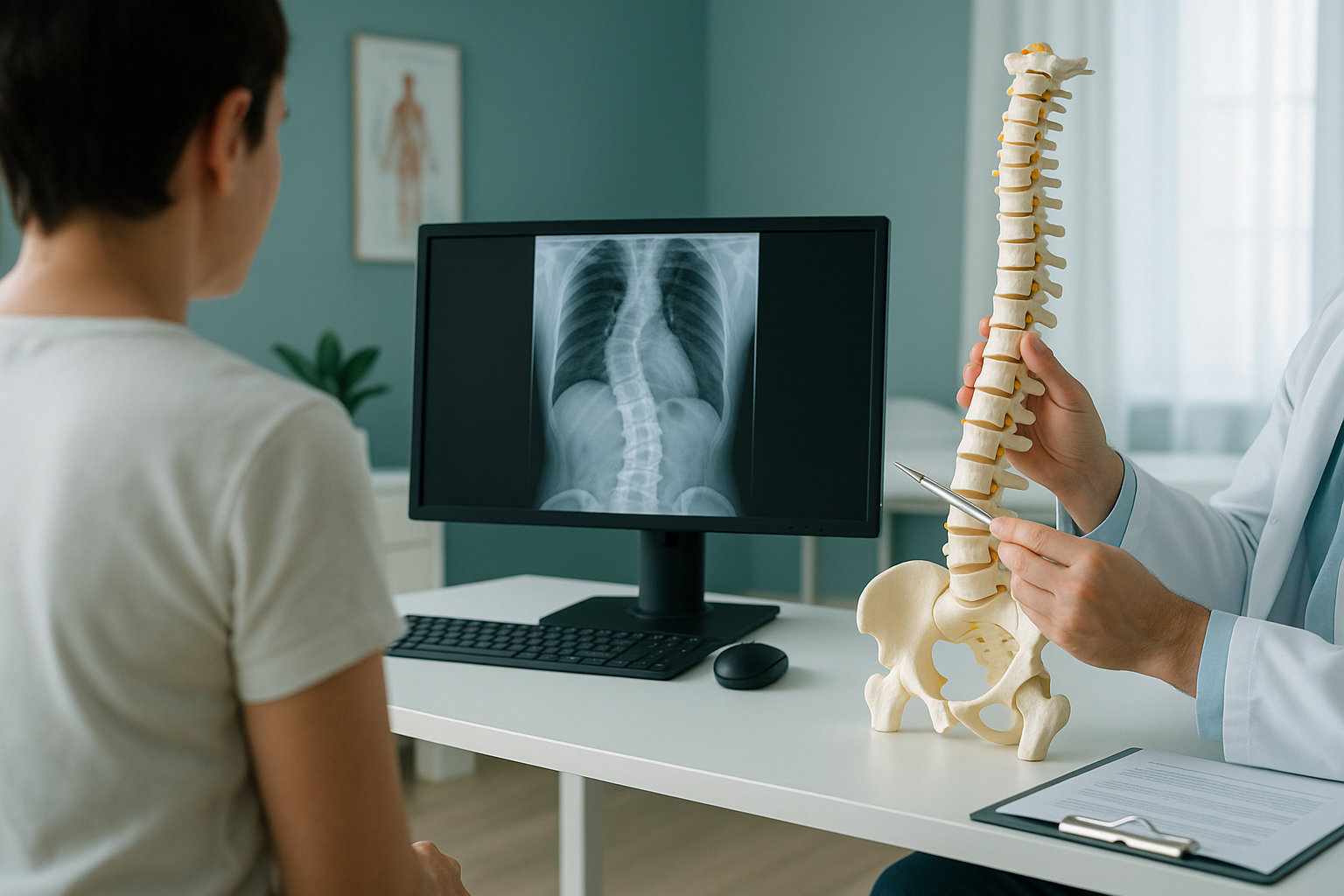
Neuromuscular scoliosis is a sideways curve of the spine caused by conditions that weaken the muscles or nerves controlling the trunk, such as cerebral palsy, muscular dystrophy, or spina bifida. Because the muscles cannot hold the spine straight, the curve often progresses steadily and can affect sitting balance, comfort, and breathing. Regular monitoring is essential.
Key takeaways
- Neuromuscular scoliosis is driven by muscle weakness or nerve problems, not by posture or habit.
- It tends to progress faster than other types of scoliosis, especially during growth spurts.
- The curve often affects the pelvis, making comfortable seated positioning difficult.
- Regular imaging surveillance helps the clinical team track progression and plan interventions at the right time.
- Low-dose imaging options can reduce cumulative radiation for patients who need many scans over their lifetime.
When the muscles around the spine are too weak or too stiff to provide balanced support, the spine gradually drifts into a curve. Unlike other forms of scoliosis, neuromuscular scoliosis is rarely about the bones themselves. It is about what is happening around them. The muscles and nerves that should hold the trunk upright are not working as expected, so gravity slowly pulls the spine off centre.
This matters because the curve does not just affect the shape of the back. It can tilt the pelvis, compress the ribs on one side, reduce lung space, and make sitting in a wheelchair painful or unstable. Understanding the condition is the first step towards getting the right monitoring and support in place.
What are the signs of neuromuscular scoliosis?
The earliest sign is often a visible lean to one side when sitting, especially without trunk support. Carers and family members usually notice changes in posture before anything else.
A noticeable lean to one side when sitting, which worsens without support.
One side of the pelvis sits higher, making level seating difficult.
Pressure on one side of the body, skin breakdown, or increasing difficulty sitting for long periods.
As the curve progresses, compressed ribs on one side may reduce respiratory function.
In many cases the person with neuromuscular scoliosis cannot describe their symptoms easily, so careful observation by carers and regular clinical review are vital.
Which conditions cause neuromuscular scoliosis?
Neuromuscular scoliosis develops when a neurological or muscular condition disrupts the normal balance of forces around the spine. The most common underlying conditions include the following.
The most common cause. Muscle stiffness or weakness on one side creates an unbalanced pull on the spine.
Progressive muscle wasting reduces trunk control, and the curve often develops once the person starts using a wheelchair full time.
Incomplete spinal development affects nerve signals below the defect, leading to asymmetric trunk support.
Spinal cord injury, spinal muscular atrophy, and other neurological conditions can also lead to neuromuscular scoliosis.
In all of these situations, the curve tends to follow a long, sweeping C-shape that often includes the pelvis. This is different from idiopathic scoliosis, which more commonly produces S-shaped curves higher up in the spine.
Why does neuromuscular scoliosis need regular monitoring?
Neuromuscular scoliosis tends to progress more quickly and more predictably than other types, particularly during growth spurts. Without regular surveillance, a moderate curve can become severe before anyone realises.
Progression matters because a worsening curve affects more than appearance. It can tilt the pelvis so far that sitting becomes impossible without specialist seating. It can compress the lungs on one side, reducing respiratory function. And it can create pressure areas that lead to skin breakdown, a serious concern for anyone who spends most of their day in a wheelchair.
The clinical team, which may include an orthopaedic surgeon, a rehabilitation specialist, a physiotherapist, and a seating engineer, relies on regular imaging to track how the curve is changing over time. The timing of interventions like bracing, seating adjustments, or surgery often depends on how fast the curve is progressing relative to remaining growth.
Thinking about a monitoring scan? If your clinical team has recommended surveillance imaging, you can discuss a monitoring scan with our radiographer in a free video call.
How is neuromuscular scoliosis imaged?
Imaging is usually done with X-ray. A full-length view of the spine, from skull to pelvis, gives the clinical team the clearest picture of the overall curve pattern and any pelvic tilt.
One challenge with standard X-rays is that they are often taken lying down, especially for people who cannot stand. But lying down removes the effect of gravity, and gravity is exactly what drives the curve in daily life. A scan taken in a supported seated or standing position can show the true shape of the spine under real-world loading.
Positioning for non-ambulant patients
For people who use a wheelchair and cannot stand unaided, positioning is one of the most important parts of the scan. At ScanAlign, low dose standing imaging, the EOS system allows imaging in a supported seated position. This means the scan can capture the spine and pelvis under gravity, closer to how the person actually sits, without requiring them to stand. Our radiographer works closely with the patient and their carer to find a comfortable, safe, and repeatable position.
Because people with neuromuscular scoliosis often need many scans over months and years, cumulative radiation exposure is a genuine concern. EOS imaging uses about 90% less radiation than a standard X-ray, around ten times less. That lower dose adds up to a meaningful difference over a lifetime of surveillance scans.
For more on how we work with younger patients and those with complex needs, see our page on paediatric imaging at ScanAlign.
A note on MRI. EOS imaging shows bones, joints, and alignment clearly. If the clinical team needs to look at soft tissue, the spinal cord, or nerve structures, MRI is usually the better tool. Your specialist will advise which type of imaging is most useful at each stage.
How is neuromuscular scoliosis managed?
Management is led by the person's wider clinical team. ScanAlign provides the diagnostic imaging that informs decisions, but does not deliver treatment. Here is a general overview of the approaches that teams commonly use.
Custom wheelchair seating, moulded supports, and sleep systems can help hold the spine in a better position and slow progression.
A spinal brace may be used to support the trunk and slow the curve, particularly in younger people who are still growing.
Stretching, strengthening, and postural programmes help maintain flexibility and comfort, even when the curve cannot be reversed.
Spinal fusion may be considered for severe or rapidly progressing curves. Detailed pre-operative imaging helps the surgical team plan the procedure.
The right combination of these approaches depends on the underlying condition, the person's age, how fast the curve is progressing, and their overall health. Accurate, repeatable imaging at each review gives the team the information they need to make well-timed decisions.
Frequently asked questions about neuromuscular scoliosis
It is a sideways curve of the spine caused by conditions that weaken the muscles or nerves controlling the trunk, such as cerebral palsy, muscular dystrophy, or spina bifida. Unlike idiopathic scoliosis, it is driven by an underlying neurological or muscular condition.
Neuromuscular scoliosis typically produces a long C-shaped curve that often involves the pelvis. It tends to progress faster and more predictably than idiopathic scoliosis, and it is more likely to affect sitting balance, respiratory function, and comfort.
In most cases, yes. The curve tends to progress, especially during periods of growth. The rate of progression depends on the underlying condition and the level of trunk muscle activity. Regular monitoring helps the clinical team intervene at the right time.
Yes. The EOS system can capture images in a supported seated position, so the person does not need to stand. Our radiographer works with the patient and their carer to find a comfortable, safe position that shows the spine under realistic loading.
An EOS scan uses about 90% less radiation than a standard X-ray, around ten times less. This is especially important for people who need repeated surveillance scans over many years.
You can self-refer. However, for complex conditions like neuromuscular scoliosis, the imaging results are most useful when shared with the wider clinical team, so we recommend discussing a scan with your consultant or GP first.
The scan itself is very quick. We allow extra time for positioning and settling, particularly for patients with complex needs. Our radiographer will discuss what to expect during the free video consultation beforehand.
The scan provides detailed information about the curve, pelvic alignment, and overall spinal balance. The decision about surgery is made by the orthopaedic or spinal team, using this imaging alongside clinical examination and other assessments.
We do not list prices on individual pages. You can find up-to-date cost information on our FAQs page.
They do different jobs. EOS shows bones, joints, and alignment under real-world loading. MRI is better for soft tissue, the spinal cord, and nerves. Your clinical team will advise which is needed at each stage.
Have questions about monitoring a neuromuscular curve?
Speak to our radiographer in a free, no-obligation video call. We can discuss positioning, what the scan involves, and whether EOS imaging is right for your situation.
Book a free video consultationWritten by Abbas Dhami (Specialist Diagnostic Radiographer)
Sources
- British Scoliosis Society, "Neuromuscular Scoliosis," patient information resources. britscoliosissoc.org.uk
- Scoliosis Research Society, "Neuromuscular Scoliosis," clinical overview. srs.org
- NHS England, "Scoliosis," guidance on diagnosis and management. nhs.uk
This page is general information and not a diagnosis.
ScanAlign is the trading name of CoreMed Solutions Ltd and operates under The Harley Street Hospital's CQC licence.