
What Are the Most Common Padel Injuries?
The most common padel injuries affect the elbow, knee, shoulder, lower back, calf and Achilles tendon. Rapid direction changes, overhead smashes and glass wall rebounds place high loads on the joints. Many padel injuries are linked to underlying alignment imbalances that only show up on a standing, weight-bearing scan.
Key takeaways
- Padel elbow (lateral epicondylitis) is the single most reported overuse injury in padel.
- Knees, shoulders, lower backs and calves are all at risk, especially in midlife players who are new to the sport.
- Your body is a connected chain. A hip or pelvis imbalance can cause pain far from its source.
- A standing whole-body scan can reveal alignment problems that lying-down imaging may miss.
- Warming up, building load gradually and addressing asymmetry are your best defences.
Which body areas are most affected by padel injuries?
Padel demands explosive side-to-side movement, sudden stops, overhead reaching and repetitive wrist action. The combination stresses several areas at once. Here are the sites that clinicians see most often.
Important. If you develop sudden numbness, weakness in a limb, or loss of bladder or bowel control after an injury, seek urgent medical attention through your GP, 111 or A&E. Do not wait for a scan.
Why does padel cause so many injuries in new players?
Padel is easy to pick up socially but physically demanding from day one. Many new players jump from little exercise to three or four sessions a week. This sudden spike in load is the biggest single risk factor for injury in recreational padel.
Midlife participants are particularly vulnerable. Tendons lose elasticity with age, and existing alignment imbalances that were never a problem in daily life can suddenly matter under match intensity.
How does whole-body alignment affect padel injuries?
Your body is a connected chain from your feet up through your pelvis, spine and shoulders. If one link is out of line, the links above and below compensate. This means the place where you feel pain is often not the place where the problem started.
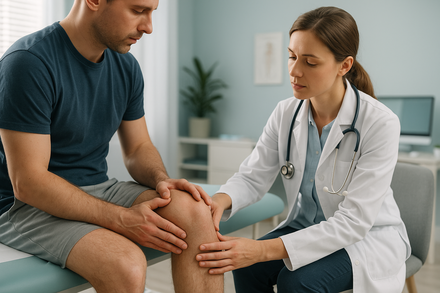
For example, a subtle pelvic tilt can change the angle at which your knee tracks during a lateral lunge. Over hundreds of direction changes per match, that small misalignment multiplies into tendon irritation or cartilage wear. Similarly, a slight spinal curve can shift shoulder load unevenly, making one rotator cuff work harder on every overhead smash.
The catch is that standard lying-down imaging rarely shows these problems. When you lie on a table for an X-ray, CT or MRI, gravity is removed, your muscles relax and your skeleton settles into a neutral position. The alignment issue that flares up on court is effectively hidden.
How can you reduce your risk of padel injuries?
You can lower your risk significantly by managing load, warming up properly, and addressing any underlying alignment issues. Read our broader guide on sports injury prevention for more detail. Here are the most important principles for padel specifically.
- Build up gradually. If you are new to padel, limit sessions to two or three per week for the first month. Your tendons need time to adapt.
- Warm up the right muscles. Lateral lunges, calf raises and gentle shoulder rotations prepare the exact movements padel demands. Five to ten minutes is enough.
- Check your grip size. A racket grip that is too small forces your forearm to overwork, accelerating padel elbow.
- Wear court-specific shoes. Running trainers lack lateral support. A proper court shoe helps protect your knees and ankles during side-to-side movement.
- Balance your body. Strengthening your non-dominant side reduces the asymmetry that one-sided racquet sports create over time.
- Listen to niggles. Persistent elbow ache, knee stiffness or back tightness after playing are early warnings. Addressing them early is far easier than managing a chronic injury.
How does a standing EOS scan help with recurring padel injuries?
A standing EOS scan captures your entire skeleton in an upright, weight-bearing position. This reveals how your bones, joints and alignment actually behave under gravity, the same conditions you experience on court.
Because the scan covers you from head to toe in one image, it can show whether a knee problem is being driven by a pelvic tilt, whether shoulder overload traces back to a spinal curve, or whether a leg-length difference is putting one side under more stress. These connections are invisible on a scan of a single body part.
EOS uses about 90% less radiation than a standard X-ray, around ten times less. That makes it a comfortable choice if you are being scanned as a precaution or for monitoring.
ScanAlign standing EOS imaging at 19 Harley Street provides this full-body standing assessment with a detailed report from a specialist radiographer. No referral is needed.
When MRI is the better choice. EOS shows bones, joints and alignment clearly. For soft-tissue injuries such as torn ligaments, disc problems or nerve compression, MRI is usually the better tool. If your clinician suspects a soft-tissue injury, they may recommend MRI instead of or alongside an alignment scan.
Dealing with a padel injury that keeps coming back? A standing alignment assessment can help identify the underlying cause.
Book a free video consultationFrequently asked questions about padel injuries
Padel is not inherently bad for your joints, but the rapid direction changes and hard court surface do place significant lateral stress on knees and ankles. Players who build up gradually and address any underlying alignment issues can enjoy the sport with a much lower injury risk.
Padel elbow is the same condition as tennis elbow, known medically as lateral epicondylitis. It causes pain on the outside of the elbow from repetitive gripping and wrist extension. The shorter padel racket and enclosed court mean more volleys per rally, which can accelerate the problem.
Padel back pain often comes from repeated twisting to reach glass wall rebounds, combined with the lumbar extension needed for overhead smashes. If you have an underlying spinal asymmetry, these movements load the lower back unevenly, increasing the risk of muscle strain and disc irritation.
Yes. A pelvic tilt or leg-length difference can change the angle at which your knee tracks during lateral lunges and stops. Over time, this creates uneven stress on the cartilage and tendons around the knee, leading to injuries that keep returning even after rest.
If pain persists for more than two to three weeks after rest, keeps coming back when you return to play, or is affecting both sides of your body differently, imaging can help identify the cause. A standing alignment scan is particularly useful for injuries that do not improve with standard treatment.
A standard X-ray typically images one body part while you are lying down. An EOS scan captures your full skeleton in a standing position, showing how gravity and weight-bearing affect your alignment. This reveals compensatory patterns across joints that a single-area scan cannot detect.
Yes. EOS uses about 90% less radiation than a standard X-ray, around ten times less. It is a very low-dose imaging method suitable for adults who need whole-body skeletal assessment.
No. ScanAlign is a private, self-pay clinic. You can self-refer by booking a free video consultation to discuss whether a scan is appropriate for your situation.
Pricing details are available on the ScanAlign FAQs and cost page.
Not necessarily. Understanding the root cause of a recurring injury often means you can modify your technique, equipment or training load rather than stopping entirely. An alignment assessment can give you and your physiotherapist or coach clear information to work with.
Find out what is behind your padel injury
A free video consultation lets you discuss your symptoms with a specialist radiographer and find out whether a standing alignment scan could help.
Book a free video consultationWritten by Abbas Dhami (Specialist Diagnostic Radiographer)
Sources
- Priego-Quesada JI, et al. "Padel: A Quantitative Overview." International Journal of Environmental Research and Public Health, 2022.
- NHS. "Tennis elbow." nhs.uk, 2023.
- Courel-Ibáñez J, et al. "Health benefits and injury patterns in padel." Science & Sports, 2019.
This page is general information and not a diagnosis.
ScanAlign is the trading name of CoreMed Solutions Ltd and operates under The Harley Street Hospital's CQC licence.