
How Accurate Are Posture Measurements, Really?
Posture measurement accuracy varies widely depending on the method used, who takes the measurement, and whether you are standing or lying down. Visual checks and phone apps are the least reliable. Calibrated, weight-bearing imaging, such as an EOS scan, gives the most repeatable, verifiable results because it captures your whole skeleton in a standardised standing position.
Key Takeaways
- Every posture measurement method has a margin of error. The question is how large that margin is.
- Skin markers, soft tissue movement and different observers are the main sources of variation.
- A measurement only matters if the change it detects is larger than the method's own error range.
- Standing, weight-bearing imaging produces more consistent and clinically relevant results than lying-down scans or visual assessments.
- Reliable baseline measurements make follow-up comparisons far more useful.
You may have had your posture assessed in a physiotherapy clinic, by a chiropractor, or even with a phone app. Each time you probably got a slightly different answer. That is not unusual. Posture is notoriously difficult to measure in a way that gives the same result twice, and understanding why helps you make better decisions about your own care.
How is posture measured in practice?
Posture is measured using a range of tools, from a simple visual inspection to full-body radiographic imaging. Each method sits on a spectrum of accuracy and repeatability.
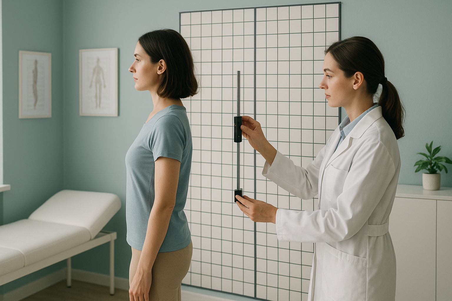
The plumb line posture test is one of the oldest clinical tools. A weighted string is hung next to you and a clinician checks whether certain landmarks line up. It is simple and free, but the result depends on where the clinician places their markers on your skin. Soft tissue, body composition, and even the time of day all influence the outcome.
Why do posture measurements vary so much?
Posture measurements vary because of a combination of observer differences, marker placement, and genuine day-to-day changes in your body. These sources of error stack on top of each other.
Intra-observer variation
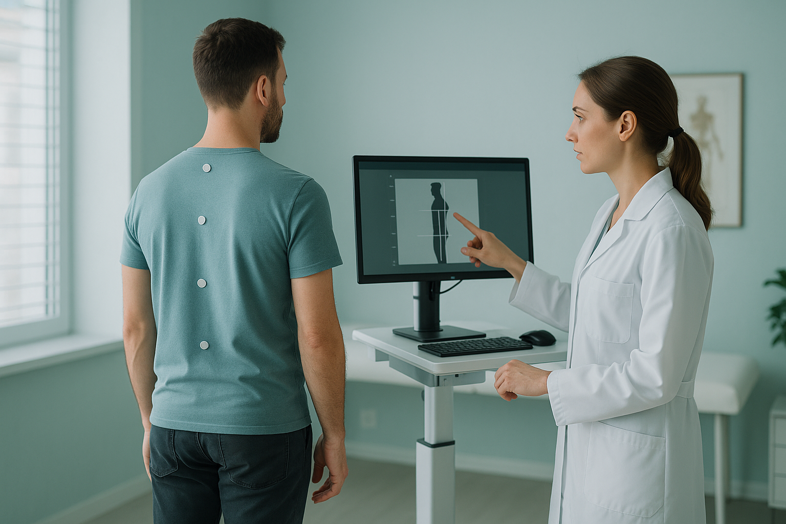
This is when the same person measures you twice and gets two different numbers. Even an experienced clinician placing skin markers on your shoulders or hips may position them slightly differently each time. That small shift changes the measurement.
Inter-observer variation
This is when two different people measure you and disagree. Studies on clinical posture assessment consistently show that agreement between different observers is only moderate, even when they follow the same protocol. In some methods, the disagreement can be large enough to make the measurement clinically meaningless.
Soft tissue and marker error
Many posture assessments rely on placing markers on the skin over bony landmarks. The problem is that skin moves. It slides over the bone beneath, especially around the pelvis and shoulders. The thicker the soft tissue, the larger the potential error. This means the marker on the outside may not represent the true position of the bone underneath.
Day-to-day variability
Your posture genuinely changes throughout the day. Disc height decreases slightly as you bear weight, muscles fatigue, and you compensate differently depending on pain levels or tiredness. A morning measurement and an afternoon measurement of the same person can produce genuinely different results, even if the measurement tool is perfect.
How does standing imaging improve posture measurement accuracy?
Standing, weight-bearing imaging improves accuracy by removing the two biggest sources of error: soft-tissue guesswork and the unnatural position of lying down.
When you lie on a table for a standard X-ray, CT or MRI, gravity is no longer pulling through your skeleton the way it does when you are upright. Your spine straightens slightly, your pelvis settles differently, and your muscles relax. The image you get is real, but it does not represent how your body actually carries load. This is why alignment problems and compensations can be missed entirely on lying-down scans.
Standardised positioning. You stand in a consistent, calibrated position. The machine, not the observer, defines the geometry. This dramatically reduces inter-observer variation.
Bone-to-bone measurement. Because the image shows your skeleton directly, there is no skin marker error. The software measures from bone landmarks, not from stickers on your skin.
Full kinetic chain. Your whole body, from feet to skull, is captured in one image. This reveals how each region relates to the next, showing compensations that single-area images miss.
Gravity removed. Lying flat removes the forces that shape your real-world posture. Curves flatten, tilts reduce, and the image may look more "normal" than your standing alignment actually is.
Single-region view. A standard X-ray or MRI typically focuses on one area. It cannot show whether your hip tilt is affecting your lower back, or whether your knee alignment is compensating for something higher up.
Still valuable for soft tissue. MRI is the gold standard for discs, nerves and soft tissue. It answers a different question to alignment imaging.
An EOS scanner is a calibrated, low-dose X-ray system that captures your full skeleton while you stand. It uses about 90% less radiation than a standard X-ray, around ten times less. The calibrated software then produces measurements directly from your bone anatomy, with high repeatability. You can learn more about how EOS scan results are reported.
For measurements like the Cobb angle measurement, which is used to assess spinal curvature, standing imaging is particularly important. The same curve can appear significantly different depending on whether you are standing under load or lying flat.
When MRI is the better tool. EOS shows bones, joints and alignment. For soft-tissue problems like disc bulges, nerve compression or ligament injuries, MRI is usually the better choice. The two often complement each other rather than compete.
What counts as a meaningful change in posture measurements?
A meaningful change is one that is larger than the measurement method's own margin of error. If the method's variability is bigger than the change it is trying to detect, the finding may not be real.
This is a critical concept. If a posture grid has, say, a natural variability of several units between assessments, then any change smaller than that variability could just be measurement noise rather than a genuine shift in your alignment. You cannot know whether you have improved or worsened unless the tool you use is precise enough to tell the difference.
Calibrated imaging systems have much tighter repeatability. That means even smaller changes between a baseline scan and a follow-up scan can be detected with confidence. This makes calibrated imaging especially valuable when tracking progress over time, for example before and after surgery, or during a rehabilitation programme.
A reliable baseline is the starting point for any meaningful follow-up. See how EOS scan results are reported to understand what your measurements mean.
Without a standardised baseline, follow-up measurements are less useful because you cannot be sure both assessments were done the same way. ScanAlign standing EOS imaging provides that standardised, repeatable foundation.
Frequently asked questions about posture measurement accuracy
A plumb line test gives a general impression of alignment, but it depends on where the observer positions the line and which landmarks they choose. Agreement between different clinicians is often only moderate, and the same clinician may get slightly different results on different days.
Phone apps can give a rough indication, but they estimate joint positions from a photo or sensor data rather than measuring bone landmarks directly. Camera angle, clothing and lighting all affect the result, so repeatability is low.
Intra-observer variation means the same person measures you twice and gets two different numbers. Inter-observer variation means two different people measure you and disagree. Both types of error exist in every measurement method, but calibrated imaging keeps both much smaller.
Lying down removes gravity from the equation. Your muscles relax, your spine settles differently, and your pelvis tilts less. The measurement you get may not reflect how your body actually loads and compensates when you are upright and weight-bearing.
An EOS scan captures your full skeleton while you stand in a standardised position. Calibrated software then measures directly from bone landmarks rather than skin markers, which gives much higher repeatability and removes the guesswork of surface-based methods.
Yes. An EOS scan uses about 90% less radiation than a standard X-ray, around ten times less. It is a quick, well-established imaging system used in hospitals worldwide.
No. ScanAlign is a self-referral clinic, so you can book directly without a GP letter. You start with a free video consultation where a specialist radiographer reviews your concerns and confirms whether a scan is appropriate for you.
Pricing details are available on our book a measured posture assessment page or through the free video consultation. We are a private, self-pay clinic with no hidden fees.
A meaningful change is one that is larger than the measurement tool's own margin of error. If the variability of the method is bigger than the change it detects, the finding could be noise rather than a genuine shift. Calibrated imaging has a much tighter margin, so smaller real changes can be detected with confidence.
Yes, provided the baseline and follow-up measurements are taken with the same standardised method. A calibrated standing scan gives a reliable reference point, so any change detected on a later scan is more likely to reflect a genuine improvement or progression rather than measurement noise.
Get measurements you can trust
A free video consultation with a specialist radiographer will help you understand whether standing whole-body imaging is right for your situation.
Book a free video consultationWritten by Abbas Dhami (Specialist Diagnostic Radiographer)
Sources
- Fortin, C. et al. (2011). "Clinical methods for quantifying body segment posture: a literature review." Disability and Rehabilitation, 33(5), 367-383.
- Illés, T. & Somoskeöy, S. (2012). "The EOS imaging system and its uses in daily orthopaedic practice." International Orthopaedics, 36(7), 1405-1411.
- NHS England. "Diagnostic imaging: guidance on clinical indications for radiographic examinations." NHS Clinical Guidelines.
This page is general information and not a diagnosis.
ScanAlign is the trading name of CoreMed Solutions Ltd and operates under The Harley Street Hospital's CQC licence.