

Understanding Your EOS Scan Results: What Does the Report Actually Tell You?
Your EOS scan results are a detailed report based on whole-body, standing images of your skeleton. A consultant radiologist reviews the images and describes how your bones, joints and overall alignment look while you are upright and weight-bearing. The report helps your clinician plan the right next steps for you.
Key takeaways
- An EOS report covers your full skeletal alignment from head to ankles, captured while you stand.
- A consultant radiologist writes the report, which your referring clinician then uses to guide treatment decisions.
- Standing imaging can reveal alignment issues that lying-down scans may miss.
- EOS uses about 90% less radiation than a standard X-ray, around ten times less.
- For soft-tissue problems such as disc or nerve issues, MRI is usually the better tool.
What does an EOS scan report show?
An EOS scan report shows the position of your bones and joints while you are standing upright, bearing your own weight. It captures your spine, pelvis, hips, knees and ankles in a single, full-length image. This means the radiologist can assess your alignment as a connected whole rather than examining one area in isolation.
Your body works as a chain. If your pelvis tilts to one side, your spine may curve to compensate, and your knee may bear extra load. A standard X-ray of just your knee would miss that bigger picture entirely. The EOS report maps out these relationships so your clinician can trace pain or dysfunction back to its actual source.
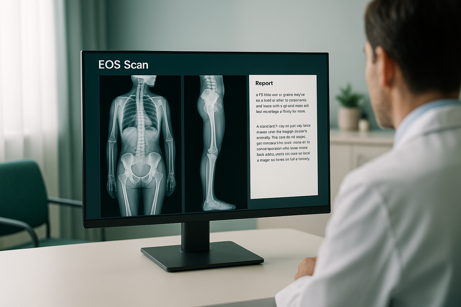
What are the key sections of an EOS scan report?
The report is structured so your clinician can quickly find the information that matters. While every report is tailored to what the images reveal, most will cover the following areas.
The report uses clinical terminology, which is why your clinician will walk you through the findings. At ScanAlign, you can discuss your results in a follow-up consultation so you understand exactly what the images mean for you.
Why do standing scan results differ from lying-down scans?
Standing imaging captures your skeleton under the natural forces of gravity and muscle tension. When you lie down for a standard X-ray, CT or MRI, your muscles relax, your spine decompresses and your pelvis settles into a neutral position. This can mask alignment problems that are clearly visible when you stand.
Think of it this way. A bridge looks different when traffic is on it compared to when it is empty. Your skeleton behaves similarly. Weight-bearing reveals how your joints cope under real-world load, not just how they look at rest.
This is why your EOS results may show findings that previous scans missed. A subtle pelvic tilt, a leg-length difference or a spinal shift may only become apparent in a standing image. Your report captures these details because the EOS system photographs you upright, from head to ankles, in one pass.
Want to understand how whole-body standing imaging works? Learn more about the EOS scan.
Who interprets EOS scan results?
A consultant radiologist reviews your EOS images and writes the formal report. Radiologists are doctors who specialise in reading medical images and identifying abnormalities. Their report is then shared with your referring clinician, whether that is an orthopaedic surgeon, a physiotherapist, an osteopath or another healthcare professional.
Your clinician interprets the report in the context of your symptoms, examination findings and medical history. This combination of expert imaging analysis and clinical knowledge ensures that any next steps are based on a complete picture.
A note on what EOS does and does not show. EOS is excellent for bones, joints and alignment. If your clinician suspects a soft-tissue problem, such as a damaged disc, a torn ligament or a trapped nerve, MRI is usually the better tool. The two types of imaging complement each other well.
What happens after you get your EOS scan results?
Once your report is ready, your clinician will review the findings with you. This conversation is where the report becomes useful. Your clinician can explain what the alignment findings mean in everyday terms and discuss whether further investigation or a referral is needed.
Common next steps include:
- Sharing the report with your surgeon ahead of a hip or knee procedure.
- Using the alignment data to guide physiotherapy or rehabilitation.
- Investigating a specific area of the spine in more detail.
- Comparing with a future scan to track changes over time.
ScanAlign is a diagnostic imaging clinic. We provide the images and the radiologist's report. We do not offer treatment, but the information we provide helps your treating clinician make better decisions.
Have questions about what your EOS scan involves, costs or radiation safety? Visit our frequently asked questions page.
Frequently asked questions about EOS scan results
It typically includes your clinical details, the radiologist's findings on bone and joint health, a full alignment assessment of your skeleton from head to feet, and a conclusion summarising the key points.
An EOS scan captures your whole body in one standing image using about 90% less radiation than a standard X-ray, around ten times less. It shows how your entire skeleton aligns under gravity, rather than imaging a single area while you lie down.
A consultant radiologist reviews the images and writes the formal report. This is a doctor who specialises in interpreting medical imaging.
No. You can self-refer. We offer a free video consultation to discuss whether an EOS scan is the right next step for your symptoms.
EOS shows bones, joints and alignment. For soft-tissue problems such as discs, ligaments or nerves, MRI is usually the better tool. The two types of imaging often work well together.
Yes. EOS uses about 90% less radiation than a standard X-ray, around ten times less. This makes it one of the lowest-dose imaging options available for skeletal assessment.
For current pricing details, please visit our FAQs and cost page.
Often, yes. Because EOS images the whole body standing up, the radiologist can identify compensatory patterns. For example, a tilted pelvis might cause pain in your lower back or knee, even though the pelvis is the root issue.
Absolutely. The report and images are yours. You can share them with any clinician, surgeon or therapist to inform treatment decisions.
If the radiologist identifies anything that requires urgent attention, your referring clinician will be informed promptly. If you experience sudden severe pain, numbness, weakness, or loss of bladder or bowel control, seek urgent medical attention through your GP, 111 or A&E immediately.
Ready to see the full picture?
A free video consultation lets you discuss your symptoms and find out whether an EOS scan is the right next step.
Book a free video consultationSources
- Illés, T. and Somoskeöy, S. (2012). The EOS imaging system and its uses in daily orthopaedic practice. International Orthopaedics, 36(7), pp.1395-1402.
- The Royal College of Radiologists. Standards for the reporting of imaging investigations (current guidance). rcr.ac.uk.
- NHS. (2023). X-ray overview, including radiation safety information. nhs.uk.
Written by Abbas Dhami (Specialist Diagnostic Radiographer).
This page is general information and not a diagnosis.
ScanAlign is the trading name of CoreMed Solutions Ltd and operates under The Harley Street Hospital's CQC licence.