
How Do Podiatrists Use Whole-Body Alignment Imaging?
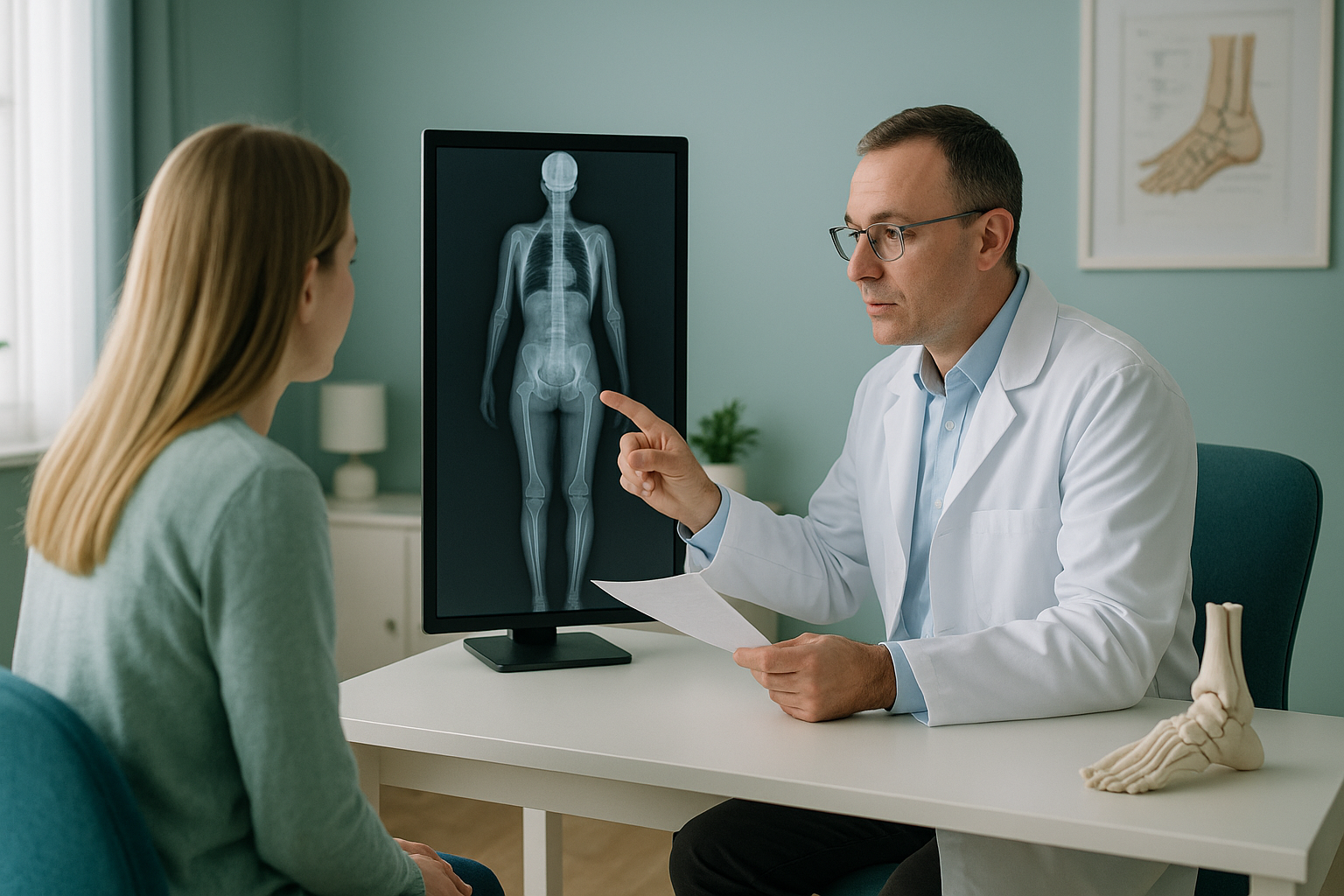
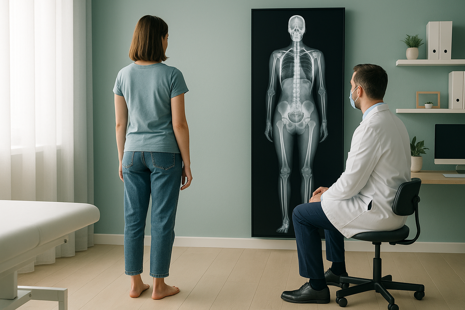
Podiatrists use whole-body standing imaging to see how the feet relate to the knees, hips, pelvis and spine under real weight-bearing load. An EOS scan captures the full kinetic chain in one low-dose exposure, giving you the alignment data you need to prescribe orthotics with greater confidence and to communicate findings clearly with other clinicians.
Key takeaways
- An EOS scan images the patient standing, capturing true load-bearing alignment from feet to head.
- It uses about 90% less radiation than a standard X-ray, around ten times less.
- Reports include full mechanical axis measurements for the lower limbs, pelvis and spine.
- No GP referral needed. You can register to refer patients directly.
- The shared report supports multidisciplinary communication with orthopaedic surgeons, physiotherapists and osteopaths.
Why does standing imaging matter for podiatry?
Standing imaging matters because the foot only functions under load. When your patient lies down for a conventional X-ray or MRI, gravity is removed. Muscles relax, arches change shape, and compensatory patterns disappear. The image you get may not reflect what the foot is actually doing during gait.
An EOS scan is taken with the patient standing naturally. This means the pronation, arch collapse or rotational asymmetry you observe in clinic is captured and measurable on the image. More importantly, you see where the foot fits into the bigger picture: the ankle, knee, hip and pelvis above it.
What does the EOS report include for podiatry referrals?
The report includes calibrated measurements of the full mechanical axis of each lower limb, pelvic obliquity, spinal balance and joint angles, all taken under standing load. For your practice, this means objective data you can use to plan orthotic prescription and to track changes over time.
Full leg alignment from hip centre to ankle, showing varus, valgus or neutral.
Whether one side of the pelvis sits higher, which can drive compensatory foot changes.
Coronal and sagittal curves, showing whether upper-body posture is affecting lower-limb loading.
True bony leg length difference, distinguished from apparent difference caused by pelvic tilt.
Which podiatry cases benefit most from alignment imaging?
Any case where foot symptoms may have a cause above the ankle benefits from seeing the whole chain. Common scenarios include persistent overpronation that does not respond to standard orthotics, recurrent plantar fasciitis, and asymmetric wear patterns that suggest a leg length difference or pelvic tilt.
Confirm whether overpronation is local or driven by knee, hip or pelvic alignment. Read more about orthotics and overpronation.
Distinguish true bony discrepancy from apparent shortening caused by pelvic obliquity or scoliosis.
Assess alignment after lower-limb surgery to guide rehabilitation and orthotic adjustments.
Evaluate rotational profiles of the tibia and femur that affect foot position and gait. Explore lower limb alignment in more detail.
How do I refer a patient to ScanAlign?
You can refer directly. No GP referral is needed. Here is the process from start to shared report.
Register as a referrer
Register to refer patients through the ScanAlign portal. It takes a few minutes and only needs to be done once.
Submit a referral
Complete the referral form with the clinical question you need answered. Tell us what you are looking for, whether that is mechanical axis, leg length, rotational profile, or a combination.
Patient attends
Your patient visits ScanAlign, whole-body standing imaging at 19 Harley Street. The scan takes around 20 minutes and uses about 90% less radiation than a standard X-ray, around ten times less.
Report shared with you
A consultant musculoskeletal radiologist reports the scan. The full report and images are shared with you and your patient, ready to inform orthotic prescription or onward referral.
Ready to start referring? Set up your account in minutes.
Register to refer patientsWhat is the difference between standing and lying-down imaging?
Lying-down imaging removes gravity from the equation. Muscles relax, joints decompress, and compensatory shifts flatten out. This means a leg length difference, pelvic tilt or rotational asymmetry may not appear on a supine scan at all.
Weight-bearing and real. The EOS system captures the patient in their natural standing posture. Gravity, muscle tone and habit all influence the image, just as they influence your patient's symptoms. Measurements reflect functional alignment, the alignment that matters when walking, standing and wearing orthotics.
Gravity removed. Conventional X-ray, CT and MRI are usually performed lying down. The spine straightens, the pelvis levels, and the feet are unloaded. These scans are excellent for identifying structural pathology, but they do not show how those structures behave under load. A patient can have a clinically significant alignment issue that simply does not appear when they are supine.
A note on MRI. EOS shows bones, joints and alignment under standing load. For soft-tissue assessment, such as tendon tears, plantar fascia thickening or nerve entrapment, MRI is usually the better tool. The two are complementary, not competing.
Which patients should I consider referring?
Not every podiatry patient needs whole-body imaging. It adds most value when you suspect the foot problem is part of a larger alignment issue, or when standard management has not produced the expected improvement.
- Persistent pronation or supination not responding to orthotics
- Suspected leg length difference
- Asymmetric symptoms or shoe wear
- Pre- or post-operative lower-limb alignment check
- Complex gait issues with suspected proximal cause
- Straightforward nail or skin conditions
- Isolated soft-tissue injury (MRI likely more useful)
- Acute trauma requiring urgent assessment
- Patients who cannot stand unsupported for 20 minutes
Frequently asked questions
Yes. ScanAlign accepts direct referrals from registered podiatrists. No GP referral is needed.
An EOS scan is a low-dose, weight-bearing X-ray that captures the whole body from head to feet in a single standing image. It uses about 90% less radiation than a standard X-ray, around ten times less.
It shows whether overpronation or other foot positions are driven by the foot itself or by alignment issues higher up, such as a pelvic tilt or knee valgus. This helps you decide whether a local orthotic is enough or whether the patient needs further assessment.
Yes. The radiation dose is about 90% less than a standard X-ray, around ten times less. It is one of the lowest-dose radiographic systems available.
ScanAlign is a private, self-pay service. Your patient pays directly. You can find pricing details on the ScanAlign FAQs page.
Yes. Once the consultant radiologist has reported the scan, the full report and images are shared with both you and your patient.
The appointment is around 20 minutes in total. The patient needs to stand still for a short period while the scan is taken.
Yes. Because the image is calibrated and taken under standing load, the report can distinguish true bony leg length difference from apparent shortening caused by pelvic tilt or spinal asymmetry.
No. EOS is a bone and joint imaging system. For soft-tissue conditions such as tendon tears, plantar fascia thickening or nerve issues, MRI is usually the better choice. The two are complementary.
Visit the referrer registration page and complete the short form. Once registered, you can submit referrals and track your patients through the portal.
Start referring your patients today
Set up your referrer account in minutes. No GP involvement needed, and your patients get the alignment data you both need.
Register to refer patients Refer a patientSources
- Illés, T. and Somoskeöy, S. (2012). The EOS imaging system and its uses in daily orthopaedic practice. International Orthopaedics, 36(7), pp.1325-1331.
- Chaibi, Y. et al. (2012). Fast 3D reconstruction of the lower limb using a parametric model and statistical inferences. Computer Methods in Biomechanics and Biomedical Engineering, 15(5), pp.457-466.
- The Royal College of Podiatry. Standards of Proficiency and Scope of Practice guidance for podiatrists in the UK.
Written by Abbas Dhami (Specialist Diagnostic Radiographer)
This page is general information and not a diagnosis.
ScanAlign is the trading name of CoreMed Solutions Ltd and operates under The Harley Street Hospital's CQC licence.