
Lower-Limb Alignment: What Is the Mechanical Axis and Why Does It Matter?
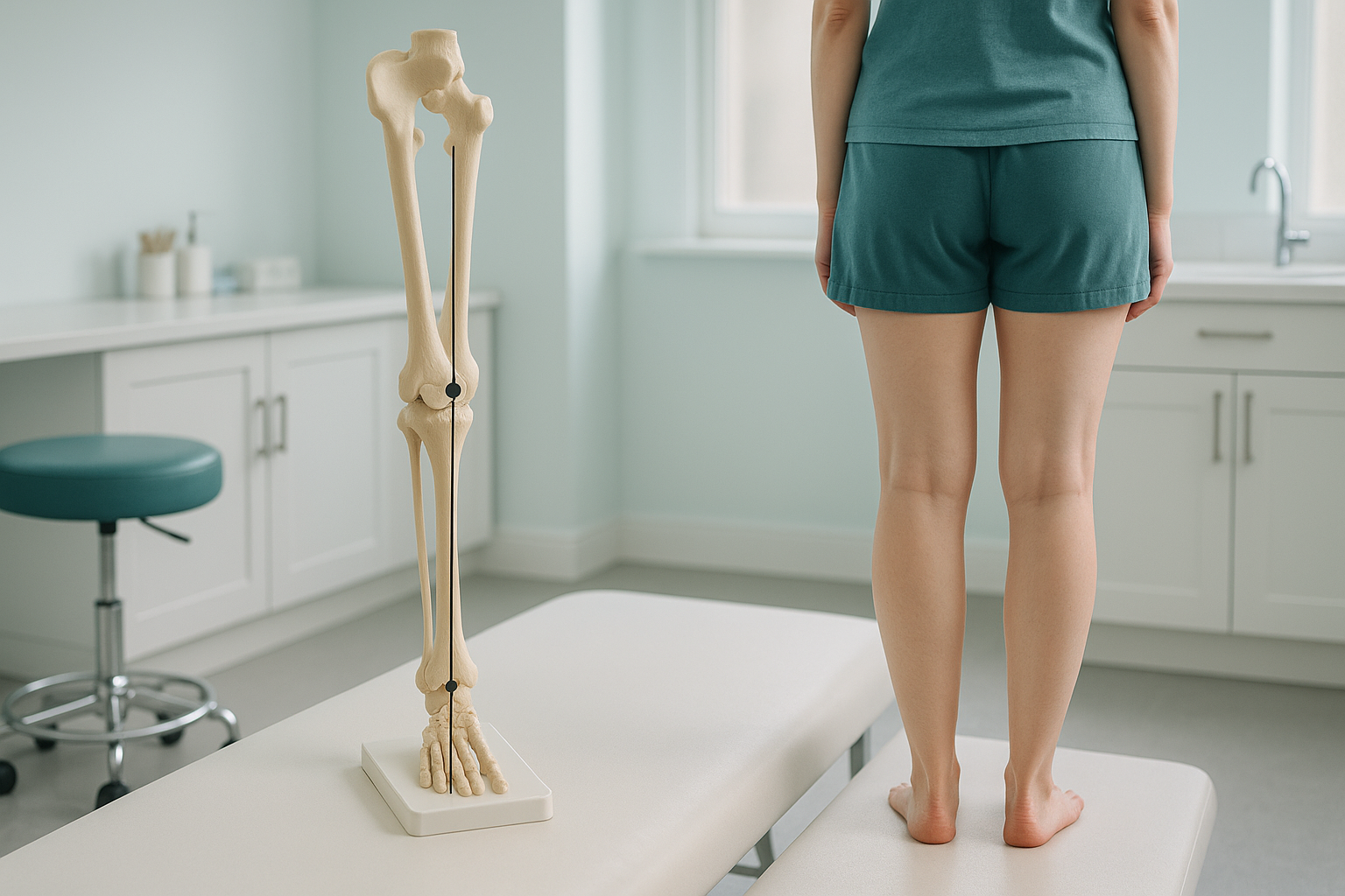
Lower-limb alignment describes how your hip, knee and ankle line up when you stand. The mechanical axis is an imaginary straight line drawn from the centre of your hip to the centre of your ankle. When that line passes through the middle of your knee, your weight is spread evenly. When it drifts to one side, extra load falls on part of the joint, which can lead to pain and wear over time.
Key takeaways
- The mechanical axis runs from hip centre to ankle centre. It should pass through the middle of the knee.
- Varus alignment (bow-legged) pushes load to the inner knee. Valgus alignment (knock-kneed) pushes it to the outer knee.
- Alignment can only be measured accurately when you are standing and weight-bearing.
- An EOS standing scan captures both legs from hip to ankle in a single low-dose image.
- Knowing your alignment helps your clinician plan the right treatment, especially before surgery.
What is the mechanical axis of the leg?
The mechanical axis is a straight line drawn from the centre of your hip joint to the centre of your ankle joint. In a normally aligned leg, this line passes through, or very close to, the centre of the knee. That means your body weight is distributed roughly equally across the inner and outer halves of the knee joint.
When the mechanical axis drifts to one side of the knee, the load is no longer shared evenly. One compartment of the knee carries more stress than the other. Over months and years, that imbalance can accelerate cartilage wear and contribute to osteoarthritis.
Varus vs valgus alignment
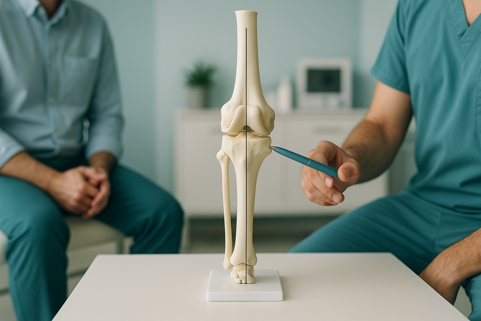
You may have heard the terms "bow-legged" and "knock-kneed." In clinical language these are called varus and valgus alignment.
- Varus (bow-legged): The mechanical axis falls to the inner side of the knee. The inside of the joint is overloaded.
- Valgus (knock-kneed): The mechanical axis falls to the outer side. The outside compartment takes more stress.
Neither is always painful. Many people live with mild varus or valgus alignment and have no problems at all. But when combined with cartilage wear, previous injury or increased activity, even a small shift in the axis can become significant.
How do you know if your leg alignment is off?
You may not notice mild malalignment on your own. Often the first clue is a pattern of symptoms that does not respond to the usual treatments.
Pain that always appears on the inner or outer side of the knee, rather than centrally.
Your shoes wear down faster on one edge, suggesting your weight is not evenly distributed.
When you stand with feet together, your knees either touch but ankles do not, or the reverse.
Physiotherapy or injections help temporarily, but the problem keeps coming back.
What causes lower-limb malalignment?
Malalignment can develop for several reasons. Sometimes it is the way your bones naturally grew. Sometimes it follows an injury or gradual wear.
Some people are born with slight curvature in the thighbone or shinbone.
As cartilage thins on one side of the knee, the joint gradually tilts.
A break near the knee that healed with a slight angle can shift the axis permanently.
Damage to supporting structures can allow the joint to drift over time.
Your body is a connected chain from feet to head. A tilt at the hip or a problem in the ankle can alter the knee's position, and vice versa. That is why a single-area scan sometimes misses the bigger picture. The pain you feel may be the result of a compensation happening somewhere else in the chain.
How is leg alignment measured when you are standing?
Accurate measurement of the mechanical axis requires a full-length image of the leg taken while you are standing and bearing your own weight. This is the only way to see how gravity and your muscles affect the position of your joints.
Conventional X-rays are usually taken lying down. When you lie flat, your muscles relax, your joints decompress, and the true alignment under load disappears. A knee X-ray taken lying down can look normal even when standing alignment is significantly off.
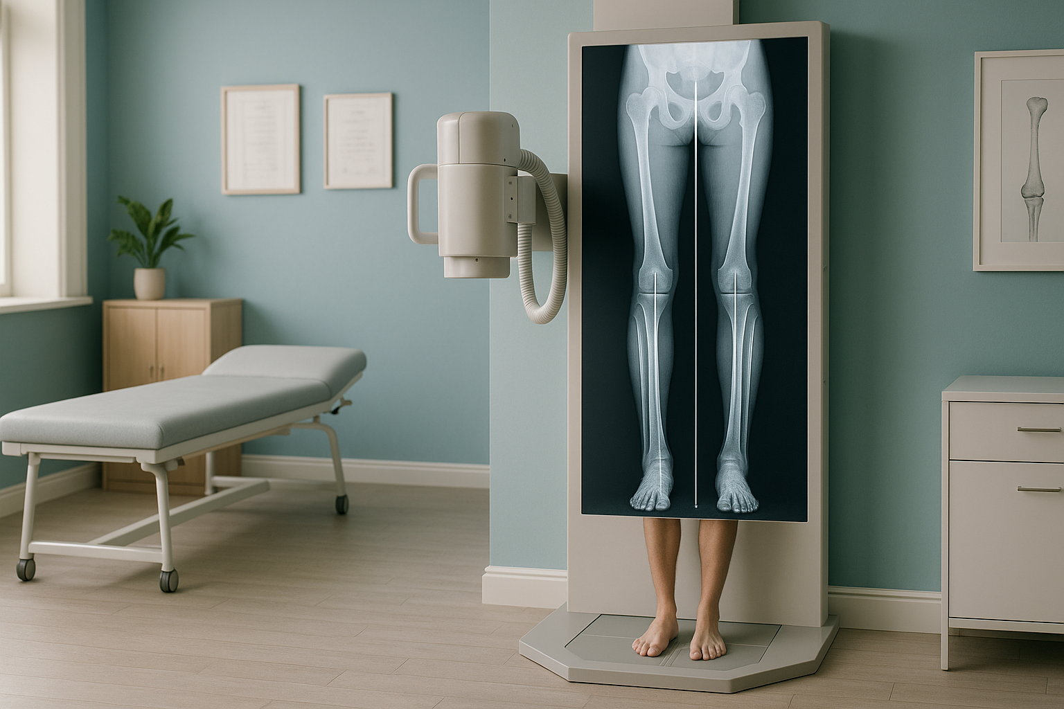
An EOS scan captures your legs from hip to ankle in a single standing image, with about 90% less radiation than a standard X-ray, around ten times less. Because the image is taken while you bear weight, it shows your joints exactly as they work in real life. The scan also captures both legs at the same time, so your clinician can compare side to side.
Want to find out whether your alignment might be contributing to your pain? A free video consultation can help you decide if an EOS scan is the right next step.
Book a free video consultationWhat can be done about lower-limb malalignment?
The right approach depends entirely on the cause and severity of the malalignment. ScanAlign is a diagnostic imaging clinic, so we provide the detailed measurements your clinician needs to make that decision. We do not deliver treatment ourselves.
In general, clinicians use alignment data to guide options such as targeted physiotherapy, orthotic insoles, or surgical planning. For example, a surgeon planning a knee replacement needs precise axis measurements to position the new joint correctly. A surgeon considering an osteotomy (a procedure that re-angles the bone) relies on the same data to calculate exactly how much correction is needed.
The most important step is getting an accurate picture of where your alignment stands today, under real weight-bearing conditions. That is what a standing full-length scan provides.
When is MRI the better choice? EOS shows bones, joints and alignment clearly. If your clinician suspects soft-tissue problems such as torn cartilage, disc issues or nerve compression, MRI is usually the better tool. Both imaging types answer different questions, and sometimes you need both.
Frequently asked questions about lower-limb alignment
In a normally aligned leg, a straight line from the centre of your hip to the centre of your ankle passes through, or very close to, the centre of your knee. Small variations are common and usually painless.
Varus means the lower leg angles inward (bow-legged), putting extra load on the inner knee. Valgus means the lower leg angles outward (knock-kneed), overloading the outer knee.
Yes. When the mechanical axis does not pass through the centre of the knee, one side of the joint takes more stress. Over time, that extra load can wear down cartilage and contribute to pain, especially during walking, running or standing for long periods.
When you lie down, gravity is no longer acting through your joints and your muscles relax. The way your bones sit together changes. Standing imaging shows how your joints work under real-life weight-bearing conditions, which is essential for accurate alignment measurement.
An EOS scan is a standing, full-body or full-leg X-ray taken in a specially designed booth. It captures both legs from hip to ankle in a single image, with about 90% less radiation than a standard X-ray, around ten times less. The result allows precise calculation of the mechanical axis and other alignment measurements. You can learn more on our EOS scan overview page.
No. ScanAlign is a private self-pay clinic and you can self-refer. We offer a free video consultation first to make sure the scan is appropriate for your situation.
The EOS system uses about 90% less radiation than a standard X-ray, around ten times less. It is one of the lowest-dose weight-bearing imaging options available. You can find more detail on our FAQs page.
Please visit our FAQs and cost page for up-to-date pricing information.
Yes. Your body works as a connected chain. A shift in the mechanical axis at the knee can change how your hip or ankle has to compensate, potentially causing pain or wear in those joints as well. That is why a full-length standing image is so valuable. You can read more on our knee alignment page.
It is critical. Your surgeon needs to know your exact mechanical axis to position the replacement joint correctly. Accurate alignment data helps restore natural load distribution and can influence how well the replacement functions long-term.
See how your legs really line up
A free video consultation with our team can help you understand whether a standing alignment scan is the right next step for you.
Book a free video consultationWritten by Abbas Dhami (Specialist Diagnostic Radiographer)
Sources
- Paley, D. Principles of Deformity Correction. Springer, 2002. (Widely referenced orthopaedic textbook on limb alignment and mechanical axis.)
- Sharma, L. et al. "The role of knee alignment in disease progression and functional decline in knee osteoarthritis." JAMA, 2001; 286(2): 188-195.
- NHS. "Knee replacement: overview." nhs.uk. (Covers the role of pre-operative alignment assessment in knee replacement planning.)
This page is general information and not a diagnosis.
ScanAlign is the trading name of CoreMed Solutions Ltd and operates under The Harley Street Hospital's CQC licence.