
At What Age Should Knock Knees in Children Correct Themselves?
Knock knees (genu valgum) are a normal part of leg development. They usually appear around age 2 to 3 and correct naturally by age 7 or 8. If a child's knees still touch with a wide ankle gap after this age, or if the angle is worsening rather than improving, a specialist assessment is a sensible next step.
Key takeaways
- Knock knees peak between ages 3 and 4, then gradually straighten.
- Most children's legs are straight or nearly straight by age 7 to 8.
- Persistent, worsening, or one-sided knock knees may need investigation.
- A standing, weight-bearing image shows leg alignment as it really is, unlike a lying-down scan.
- ScanAlign uses EOS imaging with about 90% less radiation than a standard X-ray, around ten times less.
What are knock knees in children?
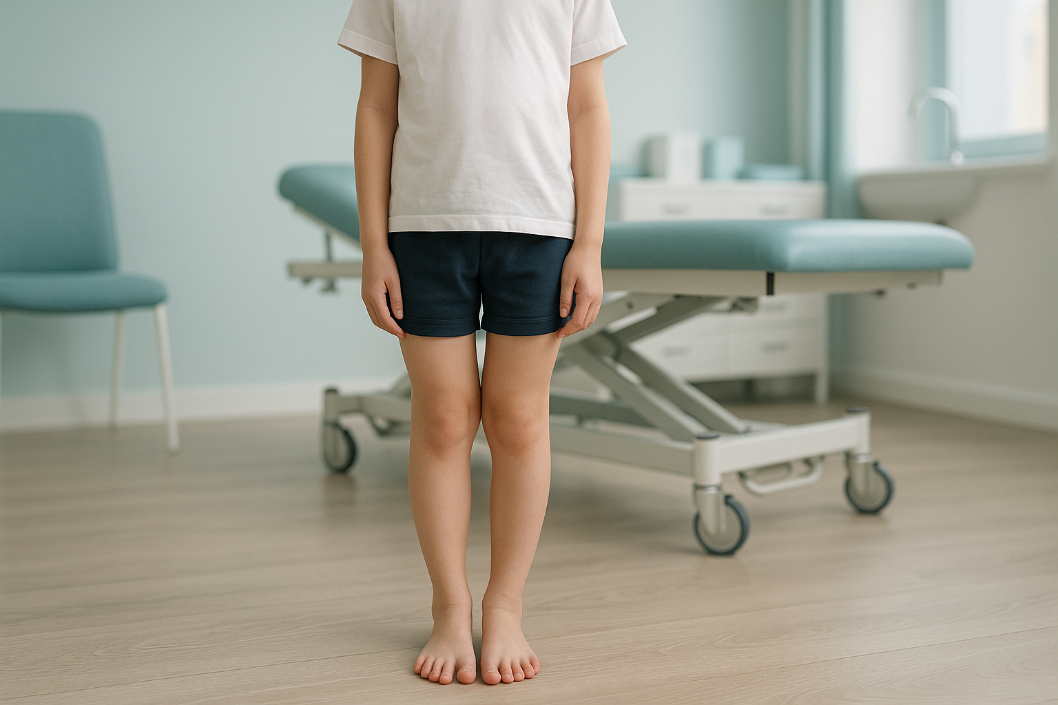
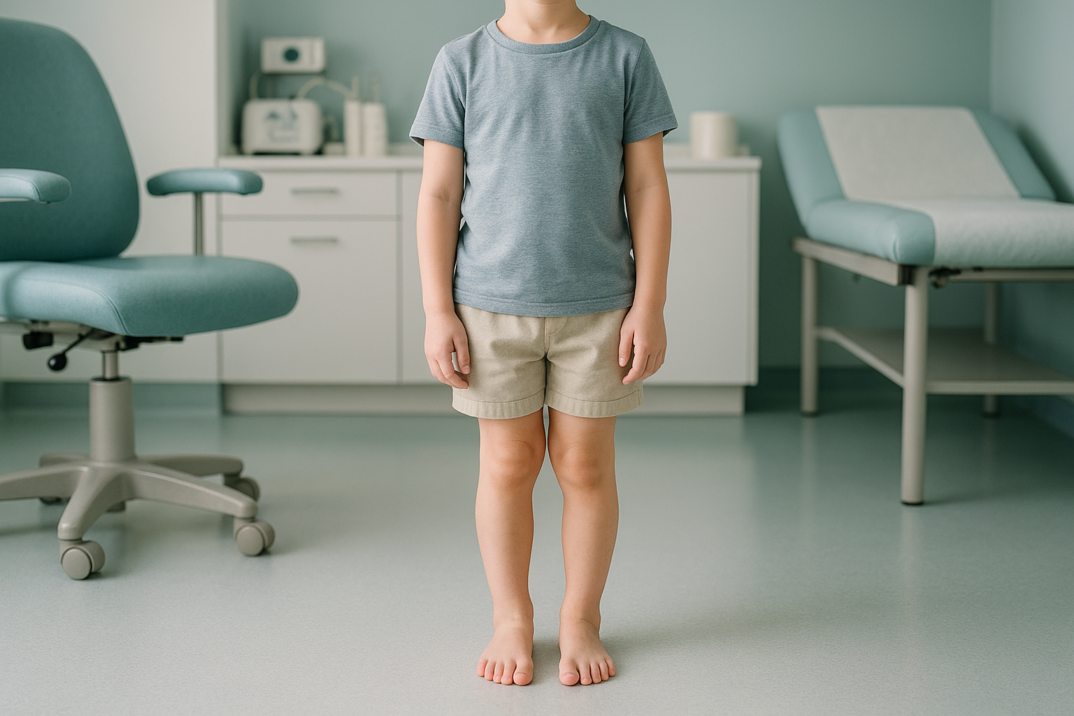
Knock knees (the medical term is genu valgum) describes a leg position where a child's knees angle inward and touch while their ankles stay apart. It looks like the legs form a wide "X" shape when your child stands up straight.
Almost every toddler goes through a knock-knee stage. It is part of the normal sequence as legs develop from the bowed shape most babies are born with through to a straighter adult alignment.
When do knock knees correct in children?
Most knock knees correct on their own by the time a child is 7 or 8 years old. The typical timeline follows a predictable pattern during normal growth.
Babies are usually slightly bow-legged. This is completely normal and relates to their position in the womb.
As a toddler grows, the legs move from bowed to a knock-knee position. This is called physiological valgus.
The inward angle is usually greatest around this age. The gap between the ankles (the intermalleolar distance) may look quite wide.
The legs gradually straighten into a slight adult valgus angle. If this correction does not happen, or if the angle worsens, further assessment is worthwhile.
Every child develops at their own pace. A slight knock-knee position that is equal on both sides and slowly improving is usually nothing to worry about.
What signs suggest knock knees may not correct on their own?
Physiological knock knees rarely cause any problems. However, certain patterns are worth checking with a specialist.
Natural correction usually finishes by this age. Persistent knock knees beyond it may indicate something other than normal growth.
If the gap between your child's ankles is widening over time rather than narrowing, this is worth investigating.
Asymmetry, where the knock knee is clearly more pronounced on one leg, may suggest a localised growth issue.
Most children with normal knock knees do not have pain. If your child is limping, falling often, or complaining of sore knees, it is worth a closer look.
What causes persistent knock knees in a child?
In most children, knock knees are simply part of normal skeletal development (physiological genu valgum). When the condition is persistent or severe, possible causes include the following.
Uneven growth at the knee growth plates can keep the inward angle going.
Low vitamin D or rickets can soften bones and contribute to a persistent valgus angle.
Rare conditions affecting bone development can present as knock knees. Early identification matters.
A fracture near the knee growth plate, even one that healed well, can change the direction of growth.
How is a child's leg alignment assessed properly?
True leg alignment is best assessed while a child is standing and bearing weight. This matters because lying down for a conventional X-ray or MRI relaxes the muscles and can change the apparent angle at the knee. The body is a connected chain from feet to head, so a tilt in the pelvis or a flat foot can push the knee inward more than it would appear on a lying-down image.
Paediatric imaging at ScanAlign uses an EOS system. EOS captures a full-length, standing image of both legs in a single exposure, with about 90% less radiation than a standard X-ray, around ten times less. Because it captures the whole limb from hip to ankle while your child stands naturally, it reveals the true mechanical alignment that a single-area image of the knee alone would miss.
When is MRI more useful? EOS shows bones, joints, and alignment clearly. If a specialist needs to look at soft tissue around the knee, such as cartilage or ligaments, MRI is usually the better tool. Both imaging types have their place.
Concerned about your child's knock knees? A short video call with our team can help you decide whether imaging is the right next step.
Book a free video consultationWhat can be done about persistent knock knees in children?
For most children, no treatment is needed at all. The knock-knee phase resolves with normal growth. When the condition is persistent or severe, a paediatric orthopaedic specialist may consider options depending on the child's age, the severity, and the underlying cause.
Possible approaches range from monitoring and observation, through to guided growth techniques in selected cases. All treatment decisions sit with the child's clinical team. ScanAlign, low dose standing imaging provides the detailed alignment images that help specialists plan the best approach, but does not deliver treatment itself.
- Knock knees are still present after age 7 or 8
- The angle seems to be getting worse
- One side looks different from the other
- Your child has knee pain, limping, or difficulty with activity
- A specialist has asked for a full-length standing leg image
- Your child is under 6 and the knock knees are mild and symmetrical
- The angle is clearly improving over time
- There is no pain, limping, or functional difficulty
If you are unsure which category your child falls into, Book a free video consultation and we will give you an honest view.
Frequently asked questions about knock knees in children
Most children's knock knees correct naturally by age 7 or 8. The inward angle peaks around ages 3 to 4 and then gradually straightens as the legs grow.
Yes. Knock knees in toddlers are a completely normal phase of leg development. After an initial bow-legged stage, most children move into a knock-knee stage around age 2 to 3.
Consider seeking advice if the knock knees persist beyond age 7 or 8, are getting worse over time, are noticeably different on each side, or are causing pain, limping, or frequent tripping.
Physiological knock knees rarely cause pain. If your child does have persistent knee pain, it is worth investigating, as it may point to an underlying cause that needs attention.
Most children do not need any treatment. The condition corrects with growth. Persistent or severe cases may be assessed by a paediatric orthopaedic specialist who can discuss options.
A specialist looks at the gap between the ankles when the knees are touching (the intermalleolar distance) and the overall mechanical axis of the leg. A standing, full-length image gives the most accurate picture.
EOS uses about 90% less radiation than a standard X-ray, around ten times less. This makes it well suited for children, where keeping radiation as low as possible is especially important.
No. ScanAlign accepts self-referrals. You can book a paediatric assessment directly through a free video consultation.
Pricing details are available on our FAQs and cost page.
Lying down relaxes the muscles and can change the way the joints sit. A standing image captures the real, weight-bearing alignment, so the clinical picture is accurate and the specialist has the most useful information.
Concerned about your child's knock knees?
A short, free video consultation with our team will help you decide whether a standing alignment scan is the right next step.
Book a free video consultationSources
- Saraph V, et al. "Physiological Genu Valgum in Children." Journal of Pediatric Orthopaedics B, 2002.
- NHS. "Knock knees." NHS Health A to Z, nhs.uk (accessed 2024).
- Stevens PM. "Guided Growth for Angular Correction." Journal of Pediatric Orthopaedics, 2007; 27(3): 253–259.
Written by Abbas Dhami (Specialist Diagnostic Radiographer)
This page is general information and not a diagnosis.
ScanAlign is the trading name of CoreMed Solutions Ltd and operates under The Harley Street Hospital's CQC licence.