
Leg Length Discrepancy in Children: How Do You Measure a Difference in a Growing Child?
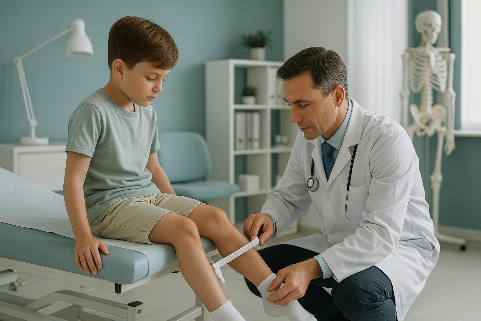
A leg length discrepancy in children is measured with a full-length, standing X-ray that captures both legs from hip to ankle in one image. Because children are still growing, a single measurement is not enough. Serial scans at regular intervals track how the difference changes over time, helping the orthopaedic team decide if and when to act.
Key takeaways
- A leg length difference in kids is common and often mild, but it can increase as the child grows.
- Accurate measurement needs a full-length standing image, not a tape measure alone.
- Serial monitoring is essential because the predicted difference at maturity guides treatment decisions.
- Each scan adds radiation, so using the lowest dose technology matters most for children.
- EOS standing imaging uses about 90% less radiation than a standard X-ray, around ten times less.
What is a leg length discrepancy in a child?
A leg length discrepancy means one leg is measurably shorter than the other. In children, this is surprisingly common. Small differences often go unnoticed. Larger ones can affect the way a child walks, cause a visible limp, or tilt the pelvis, which may lead to compensatory curves in the spine over time.
The key difference between adults and children is growth. A child's bones are still lengthening at the growth plates near the knee and hip. A small gap today can stay the same, close on its own, or widen as the child gets taller. That is why understanding the predicted difference at maturity is so important.
What are the signs your child has one leg longer than the other?
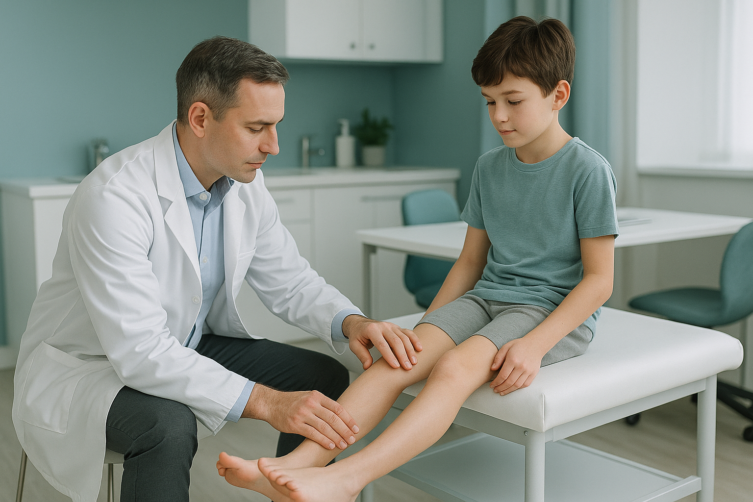
Parents usually spot a leg length difference through everyday observations rather than a formal test. Here are the most common signs.
A noticeable bob or dip when walking, especially when tired or after sport.
One side of the waistband sits higher, or a belt slopes to one side.
The child walks on tiptoe on the shorter side to level things out.
Aching after activity, sometimes on the opposite side to the shorter leg.
What causes a leg length difference in kids?
A limb length inequality in a child can be structural, meaning the bone itself is shorter, or functional, where the legs are the same length but something else, such as a tight muscle or a pelvic tilt, makes one appear shorter. The main structural causes fall into a few groups.
One growth plate grows faster or slower than its opposite, gradually widening the gap.
A break near a growth plate can speed up or slow down bone growth on that side.
Some children are born with a shorter femur or tibia on one side.
Bone infection in early childhood can disturb normal growth at the affected plate.
How is a leg length discrepancy measured accurately?
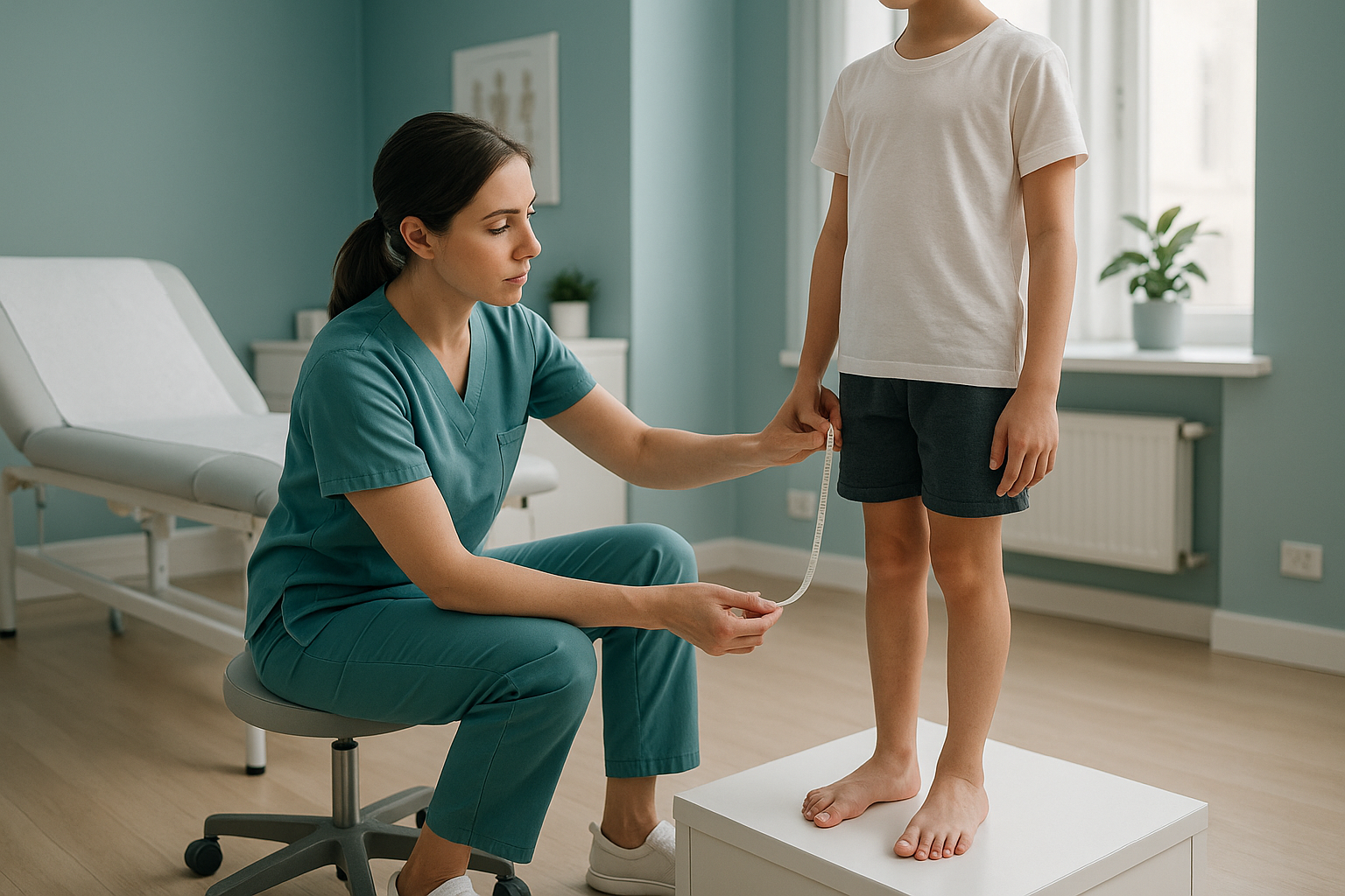
Accurate measurement requires a full-length image of both legs taken while the child is standing and weight-bearing. A tape measure from hip to ankle gives a rough guide, but it cannot separate the femur (thigh bone) from the tibia (shin bone) or reveal exactly where the shortening is. Clinicians need that detail to predict how the difference will change as the child grows.
A standing scanogram, or long-leg X-ray, captures both legs on a single image so the specialist can compare bone lengths segment by segment. When taken at intervals, usually every six to twelve months, these serial measurements build a growth curve. That curve lets the team estimate the predicted difference at maturity and decide whether a shoe raise, physiotherapy or a surgical procedure such as epiphysiodesis, where one growth plate is deliberately slowed, is the right option.
Need to start or continue a monitoring programme for your child? Paediatric imaging at ScanAlign uses ultra-low-dose EOS to track leg length over time.
Book a free video consultationWhy does standing imaging matter for a child's leg length?
When a child lies down for a conventional X-ray or CT, gravity is removed. Muscles relax, the pelvis settles flat, and any compensatory tilt the child uses in real life disappears. The image may show the bones accurately, but it misses how the child actually stands and moves.
A standing, weight-bearing image captures the full mechanical picture. It shows pelvic obliquity, any spinal curve that has developed to compensate, and the true alignment from hip to ankle under load. For the orthopaedic team, this is the information that guides real-world decisions.
Standing scan: Both legs bear weight. The pelvis tilts as it does in daily life. The image shows true leg length difference, any compensatory spinal curve, and real joint angles. This is the view the surgeon or physiotherapist needs.
Lying-down scan: Gravity is removed. The pelvis sits flat on the table. Muscles relax and may mask a functional component of the discrepancy. The bone lengths are visible, but the whole-body compensation pattern is hidden.
Learn more about EOS scanning for leg length discrepancy and how it fits into a monitoring plan.
Why does radiation dose matter most when monitoring children?
This is where leg length monitoring in children is fundamentally different from a one-off adult scan. A child who needs serial measurements may have an X-ray every six to twelve months for several years. Each scan adds to their cumulative radiation dose, and children are more sensitive to radiation than adults because their cells are dividing faster.
EOS imaging uses about 90% less radiation than a standard X-ray, around ten times less. Over a monitoring programme of, say, five or six scans across several years, that reduction adds up significantly. Less dose per scan means a far lower total exposure by the time the child reaches skeletal maturity.
At ScanAlign, low dose standing imaging, every paediatric scan is performed on the EOS system specifically because the dose question is so important for growing children.
When is MRI better? EOS shows bones, joints and alignment clearly. If the clinical team suspects a soft-tissue cause, such as a muscle contracture, a growth plate abnormality that needs cross-sectional detail, or a nerve-related issue, MRI is usually the better tool. The two scans answer different questions and are often complementary.
Frequently asked questions
Small differences are very common. Studies suggest the majority of the population has a minor limb length inequality. In most children it causes no symptoms and needs no treatment. It only becomes a clinical concern when the gap is large enough to affect gait, posture or comfort.
Sometimes. If both growth plates are healthy, the difference may stay stable or occasionally close as the child grows. In other cases the gap widens. Serial measurement is the only reliable way to find out which pattern your child follows.
Epiphysiodesis is a minor surgical procedure that slows or stops growth at one growth plate in the longer leg, allowing the shorter leg to catch up. Timing is critical and depends on the child's growth remaining, which is why accurate serial measurements are so important.
A shoe raise can level the pelvis and improve comfort while the child is growing, but it does not change bone length. It is a management tool rather than a correction. Your child's specialist will advise whether a raise is appropriate based on the size of the discrepancy.
The monitoring interval depends on the child's age, the size of the difference, and how quickly they are growing. Many specialists recommend imaging every six to twelve months during peak growth years, but your child's orthopaedic team will set the right schedule.
Yes. EOS uses about 90% less radiation than a standard X-ray, around ten times less. That lower dose is especially important for children who need repeated scans over several years, because it keeps the cumulative exposure as low as reasonably achievable.
No. ScanAlign accepts self-referred patients. You can book a paediatric leg length scan directly, and the results are shared with your child's GP or specialist.
The child stands still inside the EOS booth for about 15 to 20 seconds while the scanner moves from head to foot. There is no enclosed tunnel, no loud noise, and no injection. Most children find it straightforward.
Pricing details are on the ScanAlign FAQs and cost page. A free video consultation before booking lets you discuss whether the scan is right for your child.
Yes. Because EOS captures the whole body from head to foot in one standing image, it reveals any compensatory spinal curve, pelvic obliquity, and the leg length difference together. This gives the specialist a complete picture rather than separate snapshots of each area.
Start with a free video consultation
Talk to our team about your child's leg length concern. No referral needed, no obligation.
Book a free video consultationWritten by Abbas Dhami (Specialist Diagnostic Radiographer)
Sources
- Sabharwal S, Kumar A. Methods for assessing leg length discrepancy. Clinical Orthopaedics and Related Research, 2008; 466(12):2910-2922.
- Deschênes S et al. Diagnostic imaging in adolescent idiopathic scoliosis and leg length inequality with low-dose biplanar radiography (EOS). European Spine Journal, 2010; 19(12):2093-2101.
- Royal College of Radiologists. iRefer Guidelines: Making the Best Use of Clinical Radiology. RCR, London (current edition).
This page is general information and not a diagnosis.
ScanAlign is the trading name of CoreMed Solutions Ltd and operates under The Harley Street Hospital's CQC licence.