
How Much Radiation Is in a Scan Compared With a Flight?
A standard chest x-ray delivers about 0.02 mSv of radiation, roughly the same as a couple of hours on a long-haul flight. A CT scan of the abdomen is much higher, around 10 mSv. An EOS standing scan uses about 90% less radiation than a standard x-ray, around ten times less, making it one of the lowest-dose imaging options available.
- Millisievert (mSv) is the standard unit for measuring how much radiation your body absorbs from any source.
- We are all exposed to natural background radiation every day, about 2.2 mSv per year in the UK.
- Different scans vary enormously in dose, from a tiny fraction of background to several years' worth.
- EOS imaging uses about 90% less radiation than a standard x-ray, around ten times less.
- Context matters. The table below puts common scans side by side with everyday exposures so you can compare for yourself.
What is a millisievert and why does it matter?
A millisievert (mSv) is the unit scientists use to describe the effective dose of radiation your body receives. It accounts for both the amount of radiation and how sensitive the part of the body being exposed is. Think of it like a common currency that lets you compare very different sources, from cosmic rays at altitude to a hospital x-ray, on the same scale.
The UK average background radiation is about 2.2 mSv per year, according to Public Health England. That comes from radon gas in the ground, cosmic rays from space, and tiny amounts in the food you eat. Medical imaging is the largest man-made source of radiation for most people, which is why keeping doses as low as reasonably achievable matters.
How does the radiation from common scans compare?
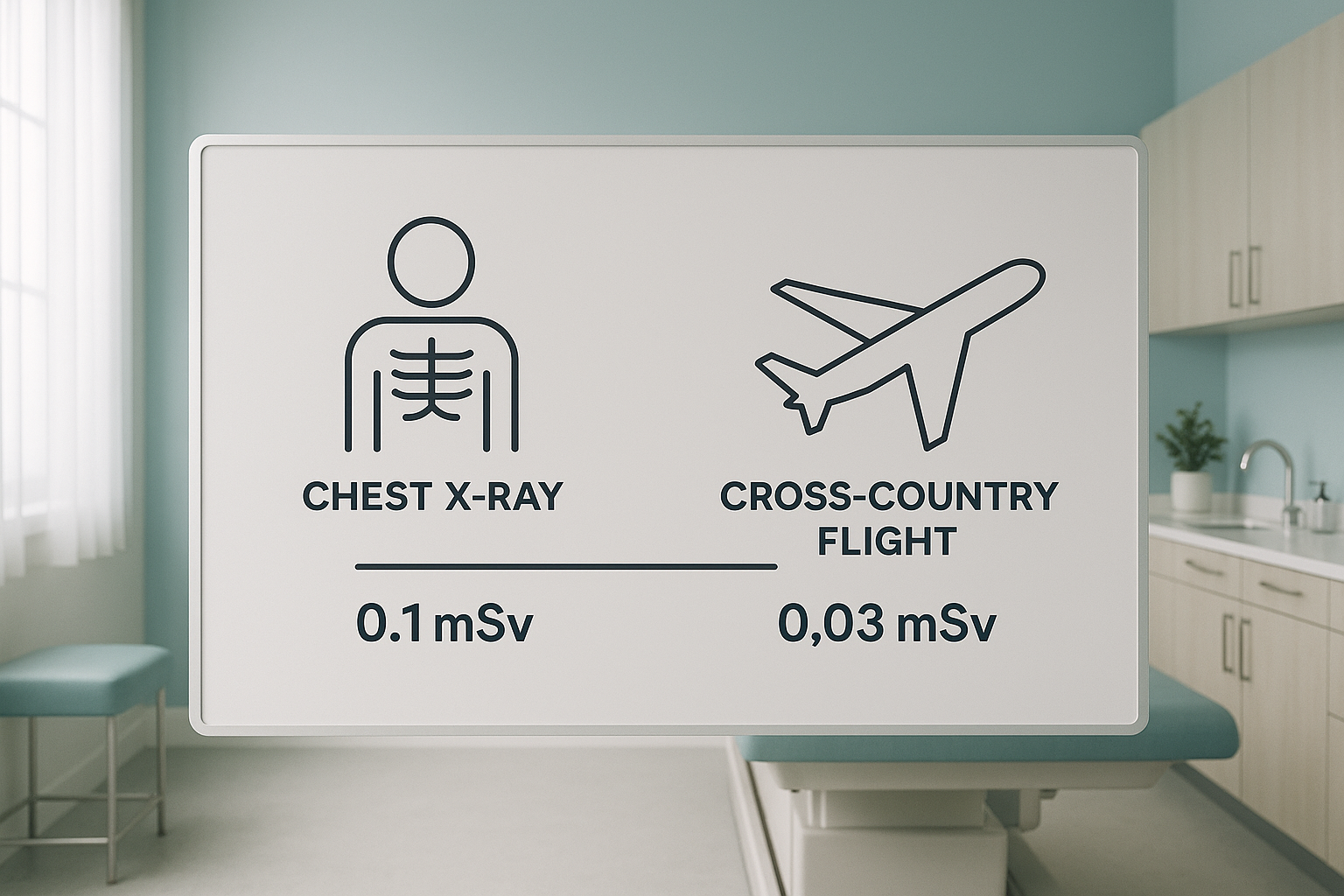
The table below puts medical imaging doses alongside everyday radiation exposures. All figures are approximate effective doses for an average adult, drawn from Public Health England (PHE) and the International Commission on Radiological Protection (ICRP).
| Source | Approximate dose (mSv) | Equivalent to | Reference |
|---|---|---|---|
| EOS standing scan | ~0.001 | A few hours of UK background radiation | EOS Imaging / manufacturer data |
| Chest x-ray | ~0.02 | About 3 days of background radiation | PHE 2022 |
| Dental x-ray (single) | ~0.005 | Less than 1 day of background | PHE 2022 |
| London to New York return flight | ~0.08 | About 2 weeks of background | PHE / ICRP |
| Lumbar spine x-ray | ~1.0 | About 5 months of background | PHE 2022 |
| CT head | ~2 | About 1 year of background | PHE 2022 |
| UK annual background (average) | ~2.2 | One year of everyday living | PHE / UKHSA |
| CT abdomen/pelvis | ~10 | About 4.5 years of background | ICRP Publication 103 |
mSv = millisievert. All figures are rounded averages for a standard adult. Actual doses vary by machine, protocol and body size.
How do you put radiation risk in context?
The risk from any single diagnostic scan is very small. To put it in perspective, the background radiation you absorb just by living in the UK for a year is about 2.2 mSv. A chest x-ray adds roughly 1% to that annual total. A transatlantic return flight adds about 4%.
Where cumulative exposure becomes worth thinking about is if you need repeated imaging over time. Each dose adds up. That is why the principle of keeping doses "as low as reasonably achievable" (known as ALARA) guides every imaging decision in the NHS and private sector alike.
For musculoskeletal imaging, where you may need follow-up scans after surgery or to track a condition like scoliosis, choosing a lower-dose option from the start can make a meaningful difference to your lifetime total. That is one of the practical advantages of EOS imaging.
Want to understand how EOS compares with other imaging types? Read our guide to EOS compared with x-ray, CT and MRI.
Why does an EOS scan use so much less radiation?
EOS technology was developed from Nobel Prize-winning particle physics research. Instead of a broad beam of x-rays (as in a conventional x-ray or CT), EOS uses a narrow fan beam that sweeps from head to toe while you stand naturally. The detector is extremely sensitive, so it needs far less radiation to produce a clear image. The result is about 90% less radiation than a standard x-ray, around ten times less.
Because you stand during the scan, the images also show your skeleton under real weight-bearing load. This matters for diagnosing alignment problems in the spine, hips and knees, where lying-down scans can relax muscles and mask the true picture. You can learn more about this on our page about EOS scan safety and radiation.
When is MRI the better choice? EOS shows bones, joints and whole-body alignment with very low radiation. For soft tissue detail such as discs, ligaments or nerves, MRI is usually the better tool because it uses no radiation at all. Your clinician will advise which scan, or combination, is right for your situation.
How do you book a low-dose standing scan at ScanAlign?
You do not need a GP referral. The process is straightforward and starts with a free video consultation so we can check that EOS is the right scan for your situation.
Free video consultation
You speak with a specialist radiographer who listens to your symptoms, reviews any previous imaging and confirms whether a standing EOS scan is appropriate.
Attend your scan
You visit ScanAlign, low dose standing imaging at 19 Harley Street. The scan takes place while you stand naturally, and is usually completed in under a minute.
Receive your report
A specialist radiologist reviews your images and produces a detailed alignment report. This is shared with you and, if you wish, your treating clinician.
Take the next step
Your report gives your clinician the diagnostic detail they need to plan treatment. ScanAlign provides the imaging, not the treatment itself.
For details on pricing and what to expect on the day, see our frequently asked questions.
Ready to see the full picture?
A free video consultation takes just a few minutes. We will let you know whether a low-dose standing scan is right for you.
Book a free video consultationFrequently asked questions about radiation dose comparison
A standard chest x-ray delivers about 0.02 mSv, which is roughly the same as 3 days of natural background radiation in the UK (source: PHE).
A return flight from London to New York exposes you to about 0.08 mSv of cosmic radiation. That is roughly equivalent to four chest x-rays.
A millisievert (mSv) is the unit used to measure effective radiation dose. It accounts for the type of radiation and the sensitivity of the body part exposed, so you can compare different sources on one scale.
An EOS scan uses about 90% less radiation than a standard x-ray, around ten times less. This makes it especially useful when repeat imaging may be needed over time.
The principle used across medicine is ALARA: "as low as reasonably achievable." No scan is done without good clinical reason. The risk from a single diagnostic scan is very small, but keeping cumulative exposure low is always sensible.
It depends on the area. A CT of the head is about 2 mSv. A CT of the abdomen and pelvis is about 10 mSv, equivalent to roughly 4.5 years of natural background radiation (source: ICRP Publication 103).
Radiation dose is cumulative over a lifetime. Each scan adds a small amount. While the risk from any single diagnostic image is very low, choosing lower-dose options when clinically appropriate helps keep your lifetime total down.
No. You can self-refer. The process starts with a free video consultation where a specialist radiographer checks that the scan is appropriate for you. You can book a low dose standing scan directly.
No. MRI uses strong magnetic fields and radio waves, not ionising radiation. It is the preferred tool when soft tissue detail (discs, nerves, ligaments) is needed. EOS is better for bones, joints and standing alignment.
Pricing information is available on our frequently asked questions page. The initial video consultation is free and carries no obligation.
Sources
- Public Health England (now UKHSA). Ionising Radiation: Dose Comparisons. GOV.UK, updated 2022. Available at: gov.uk/government/publications/ionising-radiation-dose-comparisons
- International Commission on Radiological Protection. ICRP Publication 103: The 2007 Recommendations of the International Commission on Radiological Protection. Ann ICRP 37(2-4), 2007.
- Deschênes S, Charron G, Beaudoin G, et al. Diagnostic imaging of spinal deformities: reducing patients' radiation dose with a new slot-scanning X-ray imager. Spine 2010;35(9):989-994.
Written by Abbas Dhami (Specialist Diagnostic Radiographer)
This page is general information and not a diagnosis.
ScanAlign is the trading name of CoreMed Solutions Ltd and operates under The Harley Street Hospital's CQC licence.