

What Causes Stress Fractures in Runners, and Can You Prevent Them?
A stress fracture is a tiny crack in bone caused by repetitive loading that outpaces the bone's ability to repair itself. In runners, the most common sites are the shin (tibia) and the metatarsal bones of the foot. Training load spikes, poor energy availability, low bone density and asymmetric loading from misalignment all raise the risk.
Key takeaways
- Stress fractures develop gradually from repeated impact, not a single incident.
- MRI is the first-line test for confirming an acute stress fracture.
- Alignment problems, such as leg length discrepancy, can cause asymmetric loading and repeated bone stress injuries in the same leg.
- A standing whole-body scan can reveal the biomechanical factors that standard lying-down imaging misses.
- Prevention is about load management, nutrition, bone health and correcting alignment.
If you have had more than one stress fracture, or your injuries always seem to affect the same side, the cause may not be your training plan alone. Your whole-body alignment under load, something only visible when you are standing, could be a factor that keeps getting overlooked.
What are the symptoms of a stress fracture?
The hallmark symptom is a localised, aching pain that gets worse with activity and improves with rest. Early on, the discomfort may only appear towards the end of a run. Left unchecked, it can progress until weight-bearing itself becomes painful.
Stress fracture vs shin splints. Shin splints (medial tibial stress syndrome) cause a broader, diffuse ache along the inner shin. A shin stress fracture produces sharper, pinpoint pain in one spot. Both are bone stress injuries on a spectrum, so shin splints that do not improve can sometimes progress to a stress fracture.
What causes stress fractures in runners?
Stress fractures happen when repeated loading exceeds the bone's capacity to remodel. Several factors combine to push a runner past that threshold.
When these factors overlap, the risk multiplies. A runner with a subtle leg length discrepancy who suddenly ramps up marathon training is loading one leg harder than the other, thousands of times per run. That is the recipe for a bone stress injury that keeps coming back on the same side.
How is a stress fracture diagnosed?
MRI is the first-line imaging test for a suspected acute stress fracture. It is highly sensitive, can detect bone oedema (swelling inside the bone) before a crack becomes visible on X-ray, and it shows soft-tissue detail around the injury.
When MRI is the right choice. If you suspect an active stress fracture right now, with worsening pain, point tenderness or swelling, you need an MRI. EOS shows bones, joints and alignment brilliantly, but for soft tissue, bone marrow changes and acute fracture confirmation, MRI is usually the better tool. Your GP or a sports medicine doctor can arrange one.
Plain X-rays often miss early stress fractures because the crack may not be visible for several weeks. A standard X-ray is taken lying down, which also means it cannot show how your bones align under your body weight. This matters once the acute injury has healed and you want to understand why it happened.
How does whole-body alignment affect stress fracture risk?
Your skeleton is a connected chain from your feet to your head. When one link is out of position, other areas compensate. A tilted pelvis, a difference in leg length or a rotational imbalance in the hip can redirect impact forces so that one bone absorbs far more load than it should.
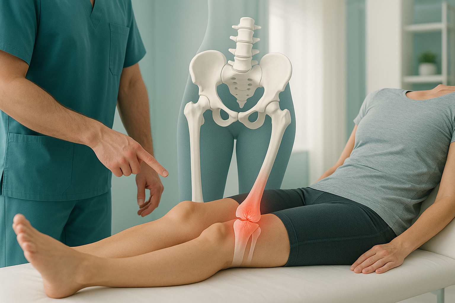
This kind of asymmetric loading only shows up when you are standing and weight-bearing. Lying-down imaging, including conventional X-ray, CT and MRI, relaxes the muscles and removes gravity. The tilt or rotation that drives uneven forces through your legs effectively disappears on the scan.
That is where ScanAlign standing EOS imaging fits in. An EOS scan captures your entire skeleton from head to feet in one upright image, using about 90% less radiation than a standard X-ray, around ten times less. It shows the alignment relationships between your pelvis, hips, knees and feet under real weight-bearing conditions, the same conditions you run in.
Had more than one stress fracture? A standing alignment assessment can help identify why your injuries keep coming back, so your physio or sports medicine specialist can address the root cause.
Book a free video consultationHow can runners prevent stress fractures?
Prevention comes down to matching the load you put through your bones with the capacity those bones have to handle it. Most advice focuses on the load side. But the alignment side, how evenly that load is distributed, is just as important for runners who keep getting injured.
For broader guidance on reducing running injury risk, read our page on sports injury prevention.
What about stress fracture recovery time?
Recovery depends on the site and severity. Most stress fractures need several weeks of reduced loading, sometimes longer for high-risk sites like the navicular bone. Your treating clinician will guide your return-to-running plan. What matters for long-term prevention is identifying the factors that caused the fracture so you can address them before you return to full training. If alignment played a role, the same forces will still be there when you start running again.
Frequently asked questions about stress fractures
The tibia (shin) is the most common site, followed by the metatarsal bones on the top of the foot. The navicular, fibula and pelvis are less common but still well documented in runners.
It is not recommended. Continuing to run on a stress fracture risks turning a tiny crack into a complete break, which takes much longer to heal and may need surgery.
Shin splints cause a broad, diffuse ache along the inner edge of the shin. A shin stress fracture produces sharp, pinpoint pain at one spot. Both are on the bone stress injury spectrum, so persistent shin splints that do not improve deserve medical attention.
Often not in the early stages. A stress fracture may take weeks to become visible on a plain X-ray. MRI is the first-line imaging test because it detects bone marrow changes before a crack appears.
EOS is not the tool for diagnosing an acute stress fracture. That is MRI's job. EOS excels at showing whole-body bone and joint alignment under weight-bearing conditions, which helps identify the biomechanical reasons a stress fracture happened, and why it might happen again.
An EOS scan uses about 90% less radiation than a standard X-ray, around ten times less. It captures a full-body standing image in a single pass.
No. ScanAlign is a private self-pay clinic, and you can book directly without a GP referral. We do recommend you share the results with your treating clinician or sports medicine specialist.
Yes, it can contribute. Even a small difference in leg length changes the way forces distribute through your lower limbs. The shorter or longer leg may absorb more impact per stride, raising the risk of a bone stress injury on that side.
Most stress fractures need several weeks of modified activity, though high-risk sites can take longer. Your doctor or physiotherapist will guide the timeline based on the location and your progress.
You can find up-to-date pricing information on our FAQs and cost page.
Keep getting injured on the same side?
A free video consultation lets you talk through your history and find out whether a standing alignment assessment could help explain why.
Book a free video consultationWritten by Abbas Dhami (Specialist Diagnostic Radiographer)
Sources
- Warden, S.J. et al. "Management and Prevention of Bone Stress Injuries in Long-Distance Runners." Journal of Orthopaedic & Sports Physical Therapy, 2014; 44(10): 749-765.
- Nattiv, A. et al. "Stress Fracture Risk Factors, Incidence, and Classification." Clinics in Sports Medicine, 2006; 25(1): 1-16.
- NHS. "Stress fractures." NHS Inform / NHS Conditions. Available at: nhs.uk.
This page is general information and not a diagnosis.
ScanAlign is the trading name of CoreMed Solutions Ltd and operates under The Harley Street Hospital's CQC licence.